3B Scientific Hip Joint with Removable Muscles, 7 part: инструкция
Раздел: Товары для здоровья
Тип:
Инструкция к 3B Scientific Hip Joint with Removable Muscles, 7 part
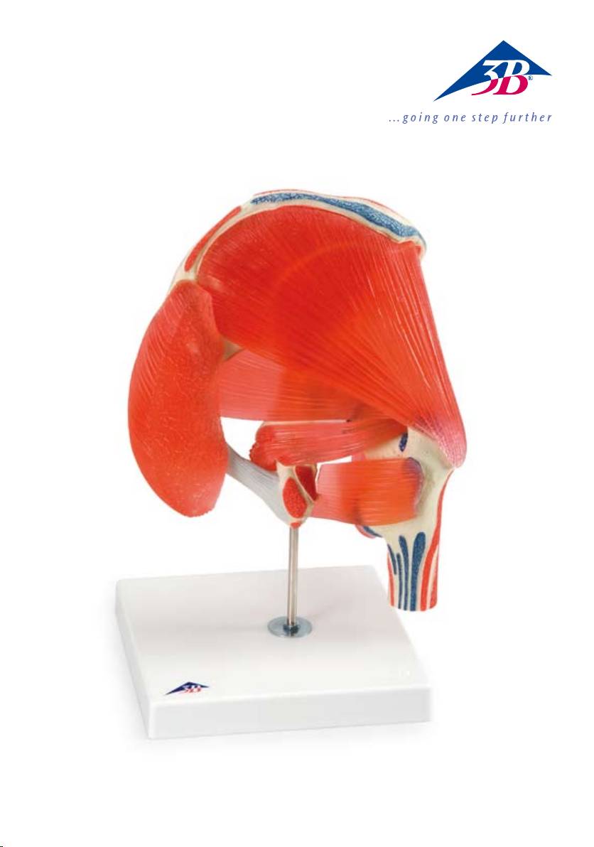
A881


®
Latin
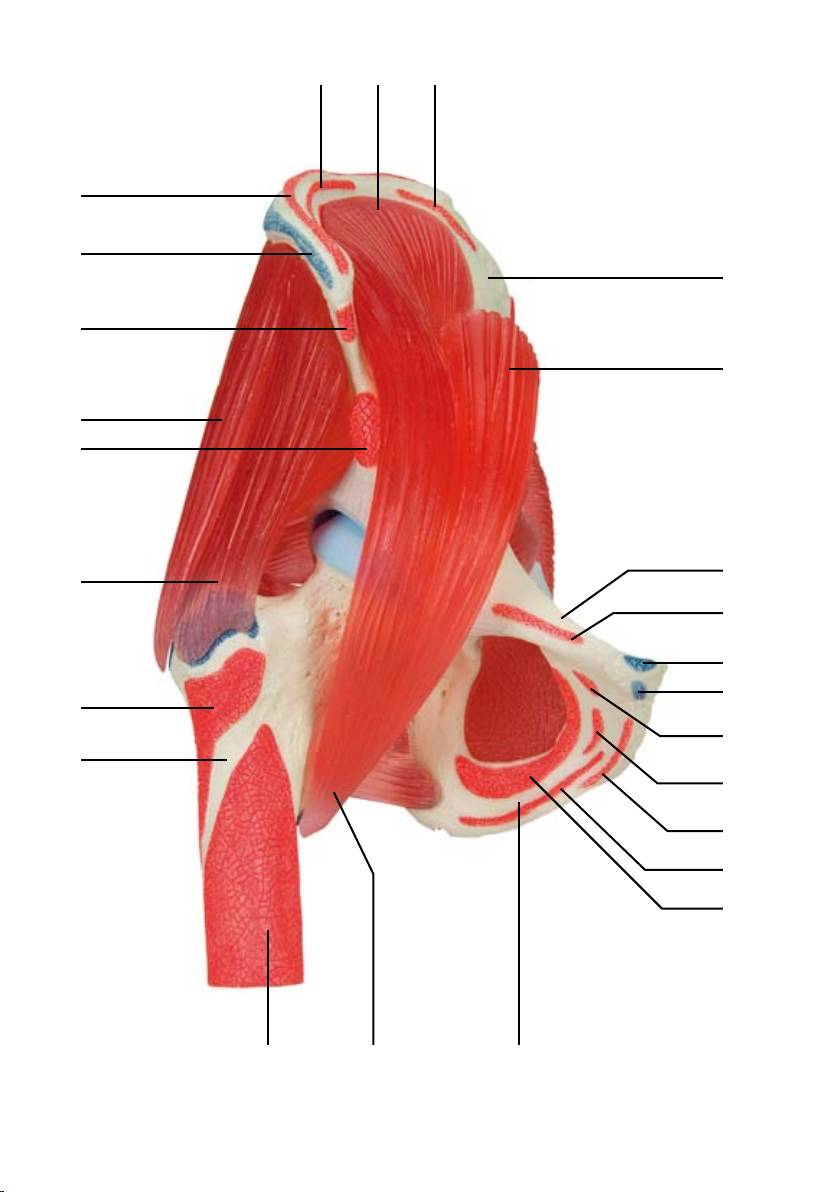
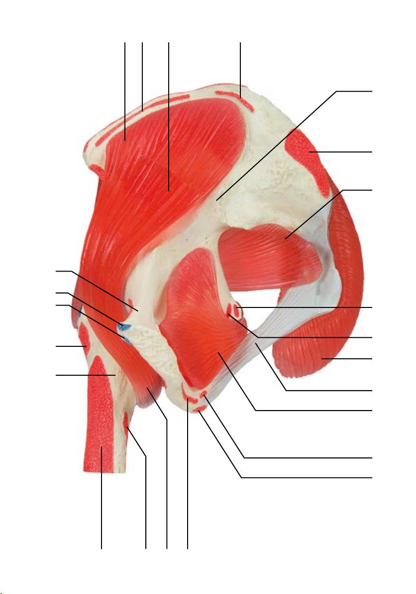
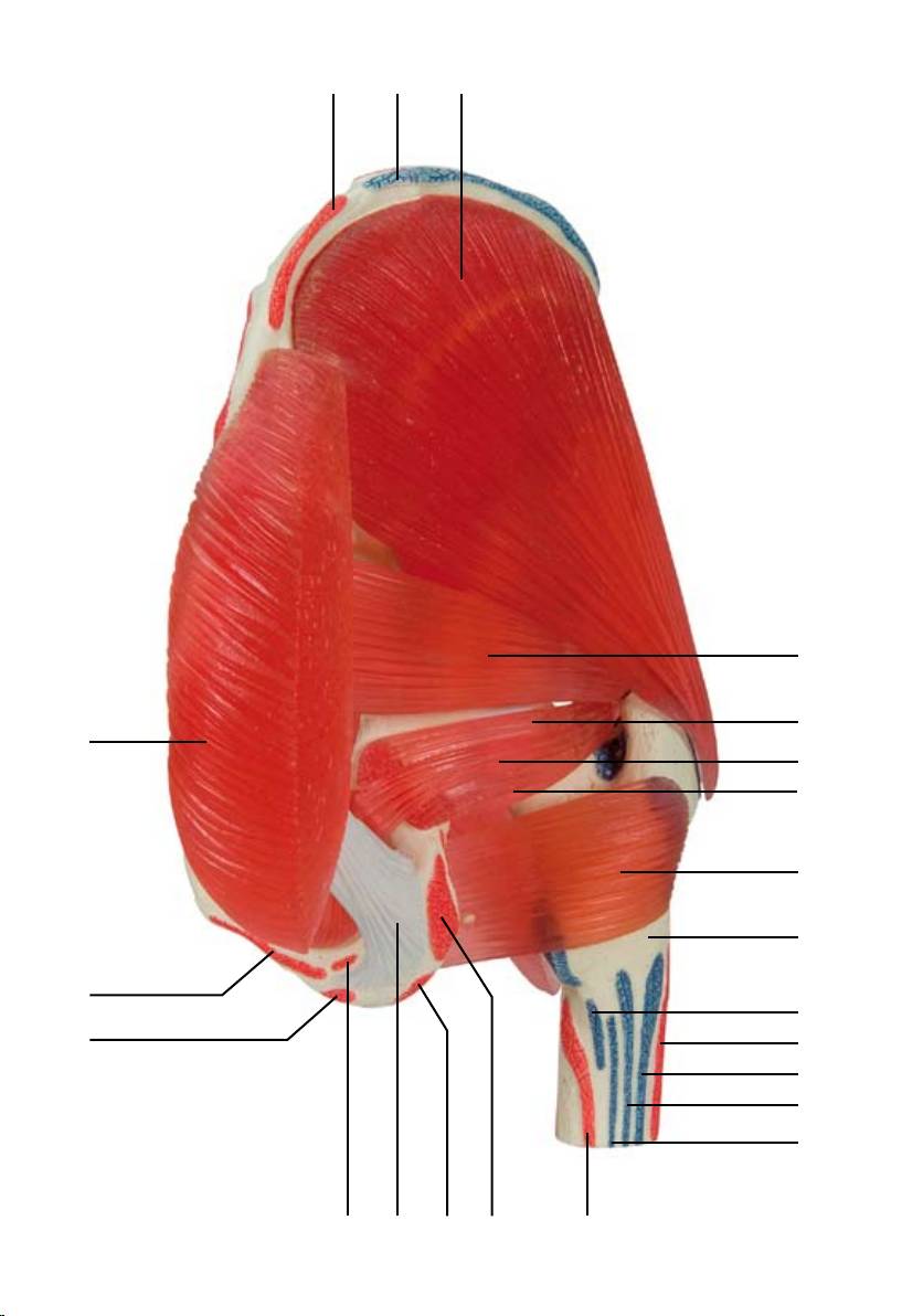
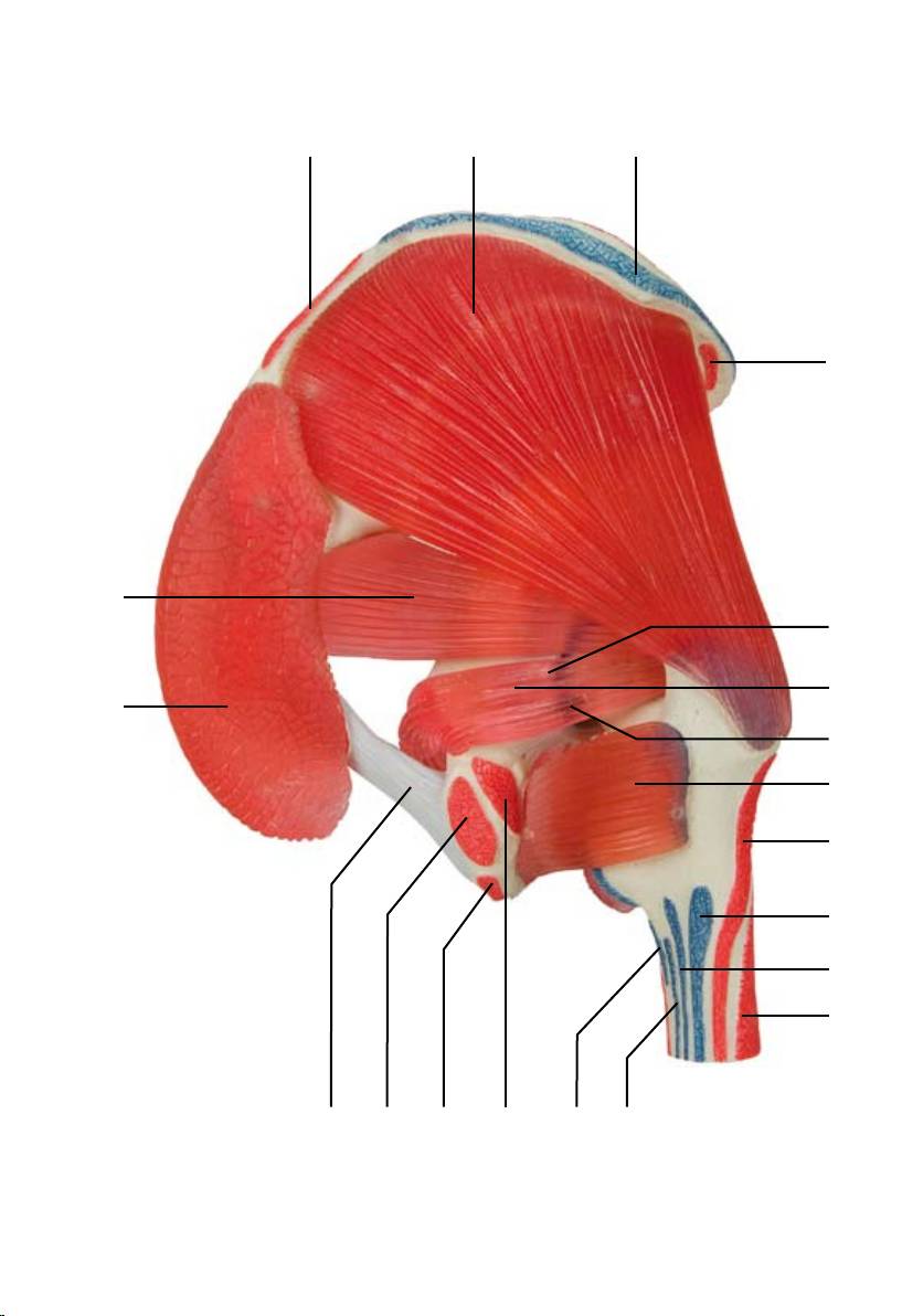
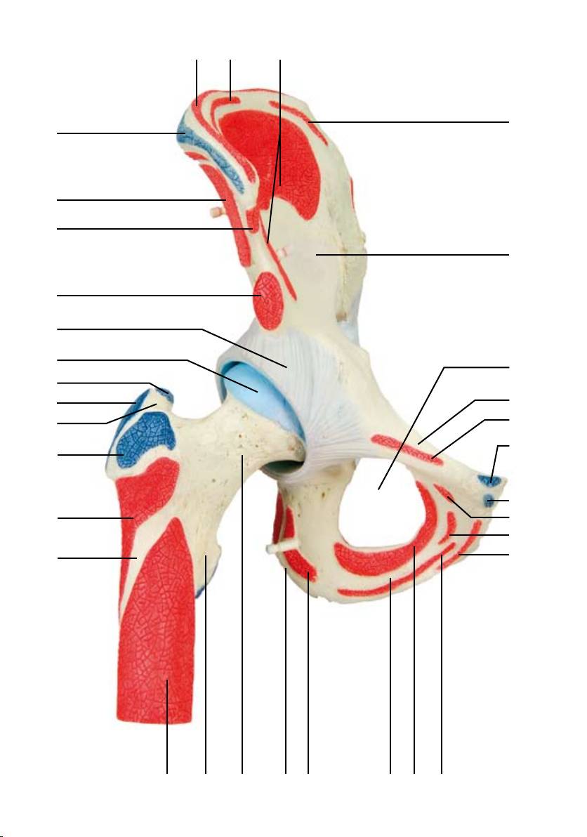
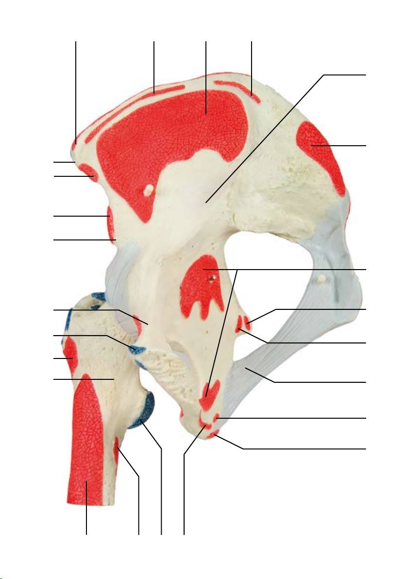
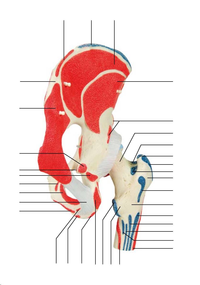
1 M. iliacus
48 M. semimembranosus, origo
2 M. quadratus lumborum, origo
49 M. iliacus, origo
3 Os coxae
50 Foramen ischiadicum majus
4 M. psoas major
51 M. quadratus femoris, origo
5 Ramus superior ossis pubis
52 Tuber ischiadicum
6 M. pectineus, origo
53 Collum femoris
7 M. rectus abdominis, insertio
54 Trochanter minor
8 M. obliquus internus abdominis, insertio
55 M. gluteus minimus, insertio
9 M. adductor longus, origo
56 Trochanter major
10 M. adductor brevis, origo
57 M. gluteus medius, insertio
11 M. gracilis, origo
58 M. piriformis, insertio
12 M. adductor magnus, origo
59 Caput femoris
13 M. obturatorius externus, origo
60 Capsula articularis
14 Ramus inferior ossis pubis
61 M. gluteus minimus, origo
15 M. iliopsoas
62 M. obturatorius internus, origo
16 M. vastus intermedius, origo
63 M. iliopsoas, insertio
17 Femur
64 Spina iliaca anterior inferior
18 M. vastus lateralis, origo
65 Spina iliaca anterior superior
19 M. gluteus minimus
66 M. gluteus medius, origo
20 M. rectus femoris, origo
67 M. obturatorius internus et mm.
21 M. gluteus medius
gemelli superior et inferior, insertio
22 M. sartorius, origo
68 M. obturatorius externus, insertio
23 M. obliquus externus abdominis, insertio
69 M. quadratus femoris, insertio
24 M. obliquus internus abdominis, origo
70 M. gemellus inferior, origo
25 M. transversus abdominis, origo
71 Spina ischiadica
26 M. erector spinae, origo
72 M. gemellus superior, origo
27 M. piriformis
73 M. gluteus maximus, origo
28 M. coccygeus, origo
29 M. levator ani, origo
30 M. gluteus maximus
31 Lig. sacrotuberale
32 M. obturatorius internus
33 M. transversus perinei superficialis, origo
34 M. ischiocavernosus, origo
35 M. transversus perinei profundus, origo
36 M. vastus medialis, origo
37 M. latissimus dorsi, origo
38 M. gemellus superior
39 M. gemellus inferior
40 M. quadratus femoris
41 M. pectineus, insertio
42 M. gluteus maximus, insertio
43 M. adductor magnus, insertio
44 M. adductor brevis, insertio
45 M. biceps femoris, origo
46 M. semitendinosus, origo
47 M. tensor fasciae latae, origo

®
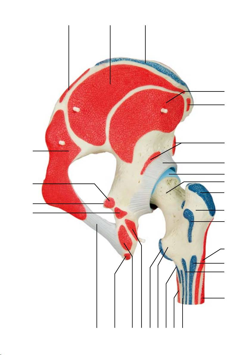
EnglishHip joint
The model shows the right hip joint of a male with the individual muscles as well as the muscle origins
and insertions on the femur and the hip bone. For educational purposes, the origin and insertion areas
of the muscles have been raised and presented in colour (muscle origin = red; muscle insertion = blue).
The hip muscles have been mounted on their corresponding regions of origin and insertion and are thus
removable.
1 Iliacus
47 Tensor fasciae latae, origin
2 Quadratus lumborum, origin
48 Semimembranosus, origin
3 Hip bone
49 Iliacus, origin
4 Psoas major
50 Greater sciatic foramen
5 Superior pubic ramus
51 Quadratus femoris, origin
6 Pectineus, origin
52 Sciatic tuber
7 Rectus abdominis, insertion
53 Neck of femur
8 Obliquus internus abdominis, insertion
54 Lesser trochanter
9 Adductor longus, origin
55 Gluteus minimus, insertion
10 Adductor brevis, origin
56 Greater trochanter
11 Gracilis, origin
57 Gluteus medius, insertion
12 Adductor magnus, origin
58 Piriformis, insertion
13 Obturator externus, origin
59 Head of femur
14 Inferior pubic ramus
60 Joint capsule
15 Iliopsoas
61 Gluteus minimus, origin
16 Vastus intermedius, origin
62 Obturator internus, oigin
17 Femur
63 Iliopsoas, insertion
18 Vastus lateralis, origin
64 Anterior inferior iliac spine
19 Gluteus minimus
65 Anterior superior iliac spine
20 Rectus femoris, origin
66 Gluteus medius,origin
21 Gluteus medius
67 Obturator internus and gemellus superior
22 Sartorius, origin
and gemellus inferior, insertion
23 Obliquus externus abdominis, insertion
68 Obturator externus, insertion
24 Obliquus internus abdominis, origin
69 Quadratus femoris, insertion
25 Transversus abdominis, origin
70 Gemellus inferior, origin
26 Erector spinae, origin
71 Ischial spine
27 Piriformis
72 Gemellus superior, origin
28 Coccygeal muscle, origin
73 Gluteus maximus, origin
29 Levator ani, origin
30 Gluteus maximus
31 Sacrotuberal ligament
32 Obturator internus
33 Superficial transverse perineal muscle,
origin
34 Ischiacavernosus origin
35 Deep transverse perineal muscle, origin
36 Vastus medialis, origin
37 Latissimus dorsi, origin
38 Gemellus superior
39 Gemellus inferior
40 Quadratus femoris
41 Pectineus, insertion
42 Gluteus maximus, insertion
43 Adductor magnus, insertion
44 Adductor brevis, insertion
45 Biceps femoris, origin
46 Semitendinosus, origin

®
Deutsch
Hüftgelenk
Das Modell zeigt das rechte Hüftgelenk eines Mannes mit einzelnen Muskeln sowie den Muskelursprüngen
und -ansätzen am Oberschenkelknochen und am Hüftbein. Aus didaktischen Gründen sind die Ursprungs-
und Ansatzflächen der Muskeln erhöht und farbig (Ursprung = rot; Ansatz = blau) dargestellt. Die
Hüftmuskulatur ist auf ihren jeweiligen Ursprungs- und Ansatzflächen aufgesteckt und somit abnehmbar.
1 Darmbeinmuskel
47 Spanner der Oberschenkelbinde, Ursprung
2 Viereckiger Lendenmuskel, Ursprung
48 Halbmembranöser Muskel, Ursprung
3 Hüftbein
49 Darmbeinmuskel, Ursprung
4 Großer Lendenmuskel
50 Großes Sitzbeinloch
5 Oberer Schambeinast
51 Viereckiger Oberschenkelmuskel, Ursprung
6 Kamm-Muskel, Ursprung
52 Sitzbeinhöcker
7 Gerader Bauchmuskel, Ansatz
53 Oberschenkelhals
8 Innerer schräger Bauchmuskel, Ansatz
54 Kleiner Rollhügel
9 Langer Schenkelanzieher, Ursprung
55 Kleiner Gesäßmuskel, Ansatz
10 Kurzer Schenkelanzieher, Ursprung
56 Großer Rollhügel
11 Schlanker Muskel, Ursprung
57 Mittlerer Gesäßmuskel, Ansatz
12 Großer Schenkelanzieher, Ursprung
58 Birnenmuskel, Ansatz
13 Äußerer Hüftlochmuskel, Ursprung
59 Oberschenkelkopf
14 Unterer Schambeinast
60 Gelenkkapsel
15 Darmbein-Lenden-Muskel
61 Kleiner Gesäßmuskel, Ursprung
16 Mittlerer Schenkelmuskel, Ursprung
62 Innerer Hüftlochmuskel, Ursprung
17 Oberschenkelknochen
63 Darmbein-Lenden-Muskel, Ansatz
18 Äußerer Schenkelmuskel, Ursprung
64 Unterer vorderer Darmbeinstachel
19 Kleiner Gesäßmuskel
65 Oberer vorderer Darmbeinstachel
20 Gerader Oberschenkelmuskel, Ursprung
66 Mittlerer Gesäßmuskel, Ursprung
21 Mittlerer Gesäßmuskel
67 Innerer Hüftlochmuskel und oberer und
22 Schneidermuskel, Ursprung
unterer Zwillingsmuskel, Ansatz
23 Äußerer schräger Bauchmuskel, Ansatz
68 Äußerer Hüftlochmuskel, Ansatz
24 Innerer schräger Bauchmuskel, Ursprung
69 Viereckiger Oberschenkelmuskel, Ansatz
25 Querer Bauchmuskel, Ursprung
70 Unterer Zwillingsmuskel, Ursprung
26 Aufrichter der Wirbelsäule, Ursprung
71 Sitzbeinstachel
27 Birnenmuskel
72 Oberer Zwillingsmuskel, Ursprung
28 Steißbeinmuskel, Ursprung
73 Großer Gesäßmuskel, Ursprung
29 Afterheber, Ursprung
30 Großer Gesäßmuskel
31 Steißbein-Sitzbein-Band
32 Innerer Hüftlochmuskel
33 Oberflächlicher querer Damm-Muskel,
Ursprung
34 Sitzbein-Schwellkörpermuskel, Ursprung
35 Tiefer querer Damm-Muskel, Ursprung
36 Innerer Schenkelmuskel, Ursprung
37 Breiter Rückenmuskel, Ursprung
38 Oberer Zwillingsmuskel
39 Unterer Zwillingsmuskel
40 Viereckiger Oberschenkelmuskel
41 Kamm-Muskel, Ansatz
42 Großer Gesäßmuskel, Ansatz
43 Großer Schenkelanzieher, Ansatz
44 Kurzer Schenkelanzieher, Ansatz
45 Zweiköpfiger Oberschenkelmuskel, Ursprung
46 Halbsehniger Muskel, Ursprung

®
Articulación de la cadera
Español
El modelo muestra la articulación de la cadera de un hombre, con músculos individuales, al igual que sus
orígenes e inserciones en el fémur y en el hueso ilíaco. Por razones didácticas, las superficies de los orí-
genes e inserciones de los músculos se presentan en relieve y con distintos colores (origen = rojo, inserción
= azul). La musculatura de la cadera se encuentra insertada en sus regiones correspondientes de origen e
inserción y es también desmontable.
1 M. ilíaco
48 M. semimembranoso, origen
2 M. cuadrado lumbar, origen
49 M. ilíaco, origen
3 Hueso coxal
50 Orificio ciático mayor
4 M. psoas mayor
51 M. cuadrado femoral, origen
5 H. pubis, rama superior
52 Tuberosidad isquiática
6 M. pectíneo, origen
53 Fémur, cuello
7 M. recto del abdomen, inserción
54 Trocánter menor
8 M. oblicuo interno del abdomen, inserción
55 M. glúteo menor, inserción
9 M. aproximador largo, origen
56 Trocánter mayor
10 M aproximador corto, origen
57 M. glúteo medio, inserción
11 M. grácil, origen
58 M. piriforme, inserción
12 M. aproximador mayor, origen
59 Fémur, cabeza
13 M. obturador externo, origen
60 Cápsula articular
14 H. pubis, rama inferior
61 M. glúteo menor, origen
15 M. iliopsoas
62 M. obturador interno, origen
16 M. vasto intermedio, origen
63 M. iliopsoas, inserción
17 Fémur
64 Espina ilíaca anterior inferior
18 M. vasto lateral, origen
65 Espina ilíaca anterior superior
19 M. glúteo menor
66 M. glúteo medio, origen
20 M. recto femoral, origen
67 Músculo obturador interno y gemelos
21 M. glúteo medio
superior e inferior, inserción
22 M. sartorio, origen
68 M. obturador externo, inserción
23 M. oblicuo externo del abdomen, inser ción
69 M. cuadrado femoral, inserción
24 M. oblicuo interno del abdomen, origen
70 M. gémino inferior, origen
25 M. transverso del abdomen, origen
71 Espina isquiática
26 M. erector de la espina dorsal, origen
72 M. gémino superior, origen
27 M. piriforme
73 M. glúteo mayor, origen
28 M. coccígeo, origen
29 M. elevador del ano, origen
30 QM. glúteo mayor
31 Lig. sacrotuberoso
32 M. obturador interno
33 M. transverso perineal superficial, ori gen
34 M. isquiocavernoso, origen
35 M. transverso perineal profundo, origen
36 M. vasto medial, origen
37 M. latísimo del dorso, origen
38 M. gémino superior
39 M. gémino inferior
40 M. cuadrado femoral
41 M. pectíneo, inserción
42 M. glúteo mayor, inserción
43 M. aproximador mayor, inserción
44 M aproximador corto, inserción
45 M. bíceps femoral, origen
46 M. semitendinoso, origen
47 M. tensor de la fascia lata, origen

®
Français
Articulation coxofémorale
Ce modèle illustre l‘articulation coxofémorale droite chez un homme ; divers muscles y sont visibles ainsi
que les zones d‘insertion musculaire distale et proximale du fémur et de l‘os iliaque. Dans des buts didac-
t
iques, les surfaces musculaires des zones distales et proximales sont mises en surélévation et colorées
différemment (insertion distale = rouge ; insertion proximale = bleu). Les divers muscles de la hanche sont
disposés sur les surfaces respectives de leurs insertions distales et proximales, ce qui permettra de les retirer.
1 Muscle iliaque
45 Muscle biceps fémoral, origine
2 Muscle carré des lombes, origine
46 Muscle semi-tendineux, origine
3 Os iliaque
47 Muscle tenseur du fascia lata, origine
4 Muscle grand psoas
48 Muscle semi-membraneux, origine
5 Os pubien, rameau supérieur
49 Muscle iliaque, origine
6 Muscle pectiné, origine
50 Grand foramen ischiatique
7 Muscle droit de l’abdomen, insertion
51 Muscle carré fémoral, origine
8 Muscle oblique interne de l’abdomen,
52 Tubérosité ischiatique
insertion
53 Fémur, col
9 Muscle long adducteur,origine
54 Petit trochanter
10 Muscle court adducteur, origine
55 Muscle petit fessier, insertion
11 Muscle gracile, origine
56 Grand trochanter
12 Muscle grand adducteur, origine
57 Muscle moyen fessier, insertion
13 Muscle obturateur externe, origine
58 Muscle piriforme, insertion
14 Os pubien, rameau inférieur
59 Fémur, tête
15 Muscle ilio-psoas
60 Capsule articulaire
16 Muscle vaste intermédiaire, origine
61 Muscle petit fessier, origine
17 Fémur
62 Muscle obturateur interne, origine
18 Muscle vaste latéral, origine
63 Muscle ilio-psoas, insertion
19 Muscle petit fessier
64 Epine iliaque antéro-inférieure
20 Muscle droit de la cuisse, origine
65 Epine iliaque antéro-supérieure
21 Muscle moyen fessier
66 Muscle moyen fessier, origine
22 Muscle sartorius, origine
67 Insertion proximale du muscle obturateur
23 Muscle oblique externe de l’abdomen,
externe, des muscles jumeaux supérieur et
insertion
inférieur
24 Muscle oblique interne de l’abdomen, origine
68 Muscle obturateur externe, insertion
25 Muscle transverse de l’abdomen, origine
69 Muscle carré fémoral, insertion
26 Muscle érecteur du rachis, origine
70 Muscle jumeau inférieur, origine
27 Muscle piriforme
71 Epine ischiatique
28 Muscle du coccyx, origine
72 Muscle jumeau supérieur, origine
29 Muscle élévateur de l’anus, origine
73 Muscle grand fessier, origine
30 Muscle grand fessier
31 Ligament sacro-tubéral
32 Muscle obturateur interne
33 Muscle transverse superficiel du périnée,
origine
34 Insertion distale du muscle ischiocaverneux
35 Muscle transverse profond du périnée, origine
36 Muscle vaste médial, origine
37 Muscle grand dorsal, origine
38 Muscle jumeau supérieur
39 Muscle jumeau inférieur
40 Muscle carré fémoral
41 Muscle pectiné, insertion
42 Muscle grand fessier, insertion
43 Muscle grand adducteur, insertion
44 Muscle court adducteur, insertion
25 1 2
24
23
3
22
4
21
20
5
19
6
7
8
18
9
17
10
11
12
13
16 15 14
1 25 4 2
3
26
27
5
7
8
28
29
18
30
17
31
32
33
34
15
353616
37
23
21
27
38
30
32
39
40
17
35
41
34
18
42
43
44
31 4633 45 36
37 21 23
27
47
27
38
32
30
39
40
18
42
43
16
31 45 46 48 41 44
2524 49
2
23
61
22
3
20
60
1
59
50
58
5
57
6
56
7
55
15
8
18
9
10
11
17
53
14 13 1251525416
24 25 49 2
3
26
65
22
20
64
62
5
28
7
29
2
18
17
31
33
34
633616
35
37 23 66
3
61
73
1
20
53
67
72
58
15
57
32
71
68
70
56
29
69
62
31
17
35
42
43
44
41
18
3433 63 54 3646
45
37 66 23
61
47
20
73
60
59
53
72
57
71
56
70
69
18
42
43
16
45 48
44364154634631

®
PortuguêsArticulação do quadril
O modelo mostra a articulação direita do quadril de um homem com cada músculo assim como as origens
dos músculos e suas inserção no osso fêmur e no colo. Por razão didática as origens e inserção dos múscu-
los estão ressaltados e em cores (origem = vermelho; inserção = azul) na sua representação. A musculatura
do quadril está inserida dentro de cada origem e inserção, e é assim removível.
1 Músculo ilíaco
47 Músculo tensor da fáscia lata, origen
2 Músculo quadrado lombar, origen
48 Músculo semimembranoso, origen
3 Osso coxal
49 Músculo ilíaco, origen
4 Músculo psoas maior
50 Forame isquiático maior
5 Osso púbis, ramo superior
51 Músculo quadrado da coxa, origen
6 Músculo pectíneo, origen
52 Tuberosidade do ísquio
7 Músculo reto do abdome, inserção
53 Colo do fêmur
7 Músculo oblíquo interno do abdome, inserção
54 Trocânter menor
9 Músculo adutor longo, origen
55 Músculo glúteo mínimo, inserção
10 Músculo adutor curto, origen
56 Trocânter maior
11 Músculo grácil, origen
57 Músculo glúteo médio, inserção
12 Músculo grande adutor, origen
58 Músculo piriforme, inserção
13 Músculo obturador externo, origen
59 Cabeça do fêmur
14 Osso púbis, ramo inferior
60 Cápsula articular
15 Músculo iliopsoas
61 Músculo glúteo mínimo, origen
16 Músculo vasto intermédio, origen
62 Músculo obturador interno, origen
17 Fêmur
63 Músculo iliopsoas, inserção
18 Músculo vasto lateral, origen
64 Espinha ilíaca ântero-inferior
19 Músculo glúteo mínimo
65 Espinha ilíaca ântero-superior
20 Músculo reto da coxa, origen
66 Músculo glúteo médio, origen
21 Músculo glúteo médio
67 Músculo obturador interno e músculos gêmeos
22 Músculo sartório, origen
superiores e inferiores, inserção
23 Músculo oblíquo externo do abdome, inserção
68 Músculo obturador externo, inserção
24 Músculo oblíquo interno do abdome, origen
69 Músculo quadrado da coxa, inserção
25 Músculo transverso do abdome, origen
70 Músculo gêmeo inferior, origen
26 Músculo eretor da espinha, origen
71 Espinha ciática
27 Músculo piriforme
72 Músculo gêmeo superior, origen
28 Músculo coccígeo, origen
73 Músculo glúteo máximo, origen
29 Músculo elevador do ânus, origen
30 Músculo glúteo máximo
31 Ligamento sacrotuberal
32 Músculo obturador interno
33 Músculo transverso superficial do períneo,
origen
34 Músculo isquiocavernoso, origen
35 M. transverso profundo do períneo, origen
36 Músculo vasto medial, origen
37 Músculo grande dorsal, origen
38 Músculo gêmeo superior
39 Músculo gêmeo inferior
40 Músculo quadrado da coxa
41 Músculo pectíneo, inserção
42 Músculo glúteo máximo, inserção
43 Músculo grande adutor, inserção
44 Músculo adutor curto, inserção
45 Músculo bíceps da coxa, origen
46 Músculo semitendinoso, origen

®
Italiano
Articolazione dell‘anca
Questo modello mostra l‘articolazione dell‘anca destra di un uomo con i singoli muscoli e con le origini e
le attaccature muscolari sul femore e sull‘osso iliaco. Per motivi didattici, le aree di origine e attaccatura
dei muscoli sono rappresentate in rilievo e di colore diverso (origini in rosso e attaccature in blu). La mus-
colatura dell‘anca si inserisce sulle rispettive aree d‘origine e attaccatura ed è quindi possibile estrarla.
1 M. iliaco
48 M. semimembranoso, origine
2 M. quadrato del lombo, origine
49 M. iliaco, origine
3 Osso iliaco
50 Foro ischiatico grande
4 M. psoas grande
51 M. quadrato del femore, origine
5 Osso del pube, branca orizzontale
52 Tubercolo ischiatico
6 M. pettineo, origine
53 Femore, collo
7 M. retto dell‘addome, attaccatura
54 Trocantere piccolo
8 M. obliquo interno dell‘addome, attaccatura
55 M. gluteo piccolo, attaccatura
9 M. adduttore lungo, origine
56 Trocantere grande
10 M. adduttore breve, origine
57 M. gluteo medio, attaccatura
11 M. gracile, origine
58 M. piriforme, attaccatura
12 M. adduttore grande, origine
59 Femore, testa
13 M. otturatore esterno, origine
60 Capsula articolare
14 Osso del pube, branca verticale
61 M. gluteo piccolo, origine
15 M. ileopsoas
62 M. otturatore interno, origine
16 Muscolo vasto mediale, origine
63 M. ileopsoas, attaccatura
17 Femore
64 Spina iliaca anteroinferiore
18 M. vasto esterno, origine
65 Spina iliaca anterosuperiore
19 M. gluteo piccolo
66 M. gluteo medio, origine
20 M. retto del femore, origine
67 M
. otturatore interno e M. gemello superiore
21 M. gluteo medio
e inferiore, attaccatura
22 M. sartorio, origine
68 M. otturatore esterno, attaccatura
23 M. obliquo esterno dell‘addome, attaccatura
69 M. quadrato del femore, attaccatura
24 M. obliquo interno dell‘addome, origine
70 M. gemello inferiore, origine
25 M. trasverso dell‘addome, origine
71 Spina ischiatica
26 M. erettore della spina, origine
72 M. gemello superiore, origine
27 M. piriforme
73 M. gluteo grande, origine
28 M. coccigeo, origine
29 M. elevatore dell‘ano, origine
30 M. gluteo grande
31 Leg. sacrotuberoso
32 M. otturatore interno
33 M. trasverso superficiale del perineo, origine
34 M. ischiocavernoso, origine
35 M. trasverso profondo del perineo, origine
36 M. vasto mediale, origine
37 M. larghissimo del dorso, origine
38 M. gemello superiore
39 M. gemello inferiore
40 M. quadrato del femore
41 M. pettineo, attaccatura
42 M. gluteo grande, attaccatura
43 M. adduttore grande, attaccatura
44 M. adduttore breve, attaccatura
45 M. bicipite del femore, origine
46 M. semitendinoso, origine
47 M. tensore della fascia lata, origine

股関節,筋付7分解モデル
日本語
このモデルは、男性の右股関節を再現しており、それぞれの筋に加え、大腿骨と寛骨の筋に「起始/停止部を
見ることができます。分かりやすいよう、筋,起始/停止部分は隆起し、起始は赤、停止は青に色分けして表
現しています。
筋はそれぞれに対応する起始/停止部のピンにはめ込んであり、取り外しが可能です。
1 腸骨筋
48 半膜様筋,起始
2 腰方形筋,起始
49 腸骨筋,起始
3 寛骨
50 大坐骨孔
4 大腰筋
51 大腿方形筋,起始
5 恥骨上枝
52 坐骨結節
6 恥骨筋,起始
53 大腿骨頚
7 腹直筋,停止
54 小転子
8 内腹斜筋,停止
55 小殿筋,停止
9 長内転筋,起始
56 大転子
10 短内転筋,起始
57 中殿筋,停止
11 薄筋,起始
58 梨状筋,停止
12 大内転筋,起始
59 大腿骨頭
13 外閉鎖筋,起始
60 関節包
14 恥骨下枝
61 小殿筋,起始
15 腸腰筋
62 内閉鎖筋,起始
16 中間広筋,起始
63 腸腰筋,停止
17 大腿骨
64 下前腸骨棘
18 外側広筋,起始
65 上前腸骨棘
19 小殿筋
66 中殿筋,起始
20 大腿直筋,起始
67 内閉鎖筋・上双子筋・下双子筋,停止
21 中殿筋
68 外閉鎖筋,停止
22 縫工筋,起始
69 大腿方形筋,停止
23 外腹斜筋,停止
70 下双子筋,起始
24 内腹斜筋,起始
71 坐骨棘
25 腹横筋,起始
72 上双子筋,起始
26 脊柱起立筋,起始
73 大殿筋,起始
27 梨状筋
28 尾骨筋,起始
29 肛門挙筋,起始
30 大殿筋
31 仙結節靱帯
32 内閉鎖筋
33 浅会陰横筋,起始
34 坐骨海綿体筋,起始
35 深会陰横筋,起始
36 内側広筋,起始
37 広背筋,起始
38 上双子筋
39 下双子筋
40 大腿方形筋
41 恥骨筋,停止
42 大殿筋,停止
43 大内転筋,停止
44 短内転筋,停止
45 大腿二頭筋,起始
46 半腱様筋,起始
47 大腿筋膜張筋,起始

®
Russian
Тазобедренныйсустав
Модельпредставляетсобойправыйтазобедренныйсуставчеловекасизображениемотдельных
мышц,атакжеместначаламышциихприкреплениявобластибедреннойиподвздошнойкостей.Из
дидактическихсоображенийместаначалаиприкреплениямыщцсделаныобъемными,атакжевыделены
цветом(началомышцы-красныйцвет;местоприкрепления-синий.Мышцыбедраустанавливаютсяв
областиместприкрепленияиначалаиявляютсятакимобразомсъемными.
1 Подвздошнаямышца
43Большая
приводящаямышца,место
2 Квадратнаямышцапоясницы,начало
прикрепления
3 Тазоваякость
4
4Короткаяприводящаямышца,место
4 Большаяпоясничнаямышца
прикрепления
5 Верхняяветвьлобковойкости
45
Двуглаваямышцабедра,начало
6 Гребенчатаямышца,начало
46Полусухожильнаямышца,начало
7 Прямаямышцаживота,местоприкрепления
47 Мышца,напрягающаяширокуюфасцию
8 Внутренняякосаямышцаживота,место
бедра,начало
прикрепления
48Полуперепончатаямышца,начало
9 Длиннаяприводящаямышцабедра,начало
49Подвздошнаямышца,начало
10 Короткаяприводящаямышцабедра,начало
50Большоеседалищноеотверстие
11 Тонкаямышца,начало
51 Квадратнаямышцабедра,начало
12 Большаяприводящаямышцабедра,начало
52Седалищныйбугор
13 Наружнаязапирательнаямышца,начало
53Шейкабедреннойкости
14 Нижняяветвьлобковойкости
54Малыйвертел
15 Подвздошно-поясничнаямышца
55Малаяягодичнаямышца,местоприкрепления
16 Промежуточнаяширокаямышцабедра,
56Большойвертел
начало
57 Средняяягодичнаямышца,местоприкрепления
1
7 Бедреннаякость
58Грушевиднаямышца,местоприкрепления
18 Латеральнаяширокаямышцабедра,начало
59Головкабедреннойкости
19 Малаяягодичнаямышца
60Суставнаякапсула
20Прямаямышцабедра,начало
61 Малаяягодичнаямышца,начало
21 Средняяягодичнаямышца
62Внутренняязапирательнаямышца,начало
22Портняжнаямышца,начало
63Подвздошно-поясничнаямышца,место
23Наружнаякосаямышцаживота,место
прикрепления
прикрепления
6
4Нижняяпередняяподвздошнаяость
2
4 Внутренняякосаямышцаживота,начало
65Верхняяпередняяподвздошнаяость
25Поперечнаямышцаживота,начало
66Средняяягодичнаямышца,начало
26Мышца,выпрямляющаяпозвоночник,начало
67 Внутренняязапирательнаямышцаиверхняя
27Грушевиднаямыщца
инижняяблизнецовыемышцы,место
28Копчиковаямышца,начало
прикрепления
29Мышца,поднимающаязаднийпроход,начало
68Наружная
запирательнаямышца,место
30Большаяягодичнаямышца
прикрепления
31 Крестцово-бугорнаясвязка
69
Квадратнаямышцабедра,местоприкрепления
32Внутренняязапирательнаямышца
70Нижняяблизнецоваямышца,начало
33Поверхностнаяпоперечнаямышца
71 Седалищнаяость
промежности,начало
72Верхняяблизнецоваямышца,начало
34Седалищно-пещеристаямышца,начало
73Большаяягодичнаямышца,начало
35Глубокаяпоперечнаямышцапромежности,
начало
36
Медиальнаяширокаямышцабедра,начало
37 Широчайшаямышцаспины,начало
38Верхняяблизнецоваямышца
39Нижняяблизнецоваямышца
40Квадратнаямышцабедра
41 Гребенчатаямышца,местоприкрепления
42Большаяягодичнаямышца,место
прикрепления

®
髋关节 英文
模型显示男性右髋关节的各块肌肉和肌肉在股骨和髋关节的起、止端。为了方便教育,肌肉的起、
止部位采用突起和不同的颜色(起始端:红色;终止端:蓝色)。髋关节肌肉被安装在相应的起、
止部位,因此是可移除的。
1 髂肌
48 半膜肌,起端
2 腰方肌,起端
49 髂肌,起端
3 髋骨
50 坐骨大孔
4 腰大肌
51 股方肌,起端
5 上耻骨支
52 坐骨结节
6 耻骨肌,起端
53 股骨颈
7 腹直肌,插入
54 小转子
8 腹内斜肌,插入
55 臀小肌,插入
9 长收肌,起端
56 大转子
10 短收肌,起端
57 臀中肌,插入
11 股薄肌,起端
58 梨状肌,插入
12 大收肌,起端
59 股骨头
13 闭孔外肌,起端
60 关节囊
14 下耻骨支
61 臀小肌,起端
15 骼腰肌
62 闭孔内肌,起端
16 股中间肌,起端
63 骼腰肌,插入
17 股骨
64 骼前下棘
18 股外肌,起端
65 骼前上棘
19 臀小肌
66 臀中肌,起端
20 股直肌,起端
67 闭孔内肌和上孖肌和下孖肌止端
21 股中肌
68 闭孔外肌,起端
22 缝匠肌,起端
69 股方肌,起端
23 腹外斜肌,插入
70 下孖肌,起端
24 腹内斜肌,起端
71 坐骨棘
25 腹横肌,起端
72 上孖肌,起端
26 竖脊肌,起端
73 臀大肌,起端
27 梨状肌
28 尾骨肌,起端
29 肛提肌,起端
30 臀大肌
31 骶结节韧带
32 闭孔内肌
33 浅表横断会阴肌,起端
34 多孔坐骨,起端
35 深层横断会阴肌,起端
36 股内肌,起端
37 背阔肌,起端
38 上孖肌
39 下孖肌
40 股方肌
41 耻骨肌,插入
42 臀大肌,插入
43 大收肌,插入
44 短收肌,插入
45 股二头肌,起端
46 半腱肌,起端
47 阔筋膜长肌,起端

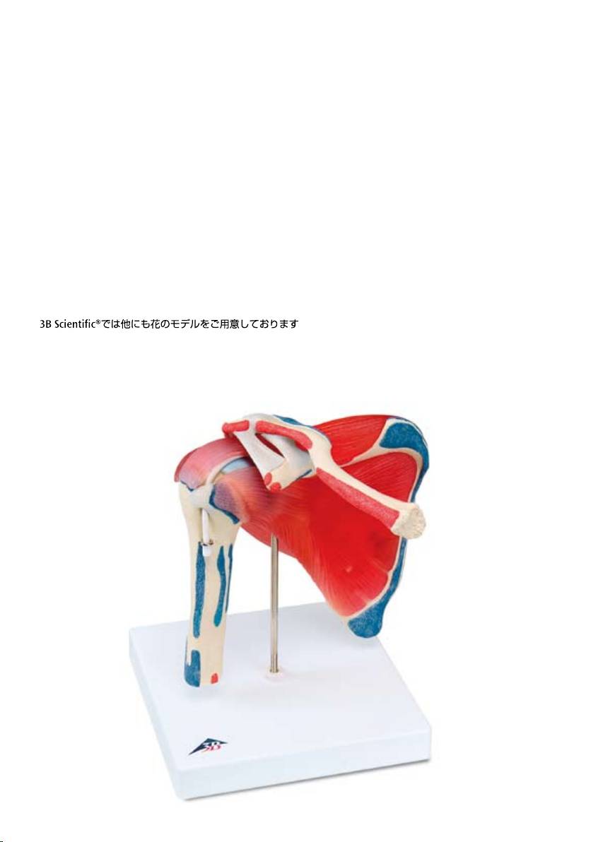
Also available from 3B Scientific®:
A880 Shoulder Joint with Rotator Cuff
Ebenf
alls bei 3B Scientific® erhältlich:
A880 Schultergelenk mit Rotatorenmanschette
T
ambien disponible en 3B Scientific®:
A880 Articulación del hombro y manguito rotatorio
Eg
alement disponible auprès de 3B Scientific® :
A880 Articulation de l‘épaule avec manchette des rotateurs
T
ambém disponível na 3B Scientific®:
A880 Articulação do ombro com manga de rotores
Disponibile
anche presso 3B Scientific®:
A880 Articolazione della spalla con Cuffia dei rotatori
A880
A880
ローテーターカフ付肩関節モデル
Also available from 3B Scientific®:
A883
Ebenf
alls bei 3B Scientific® erhältlich:
A883
T
ambien disponible en 3B Scientific®:
A883
Eg
alement disponible auprès de 3B Scientific® :
A883
T
ambém disponível na 3B Scientific®:
A883
Disponibile
anche presso 3B Scientific®:
A883
A883
A883

A881-09/06-1
© Copyright 2006 for instruction manual and design of product:
3B Scientific GmbH, Germany
®
3B S C I E NT I F I C
P R O D U C T S
3B Scientific GmbH
Rudorffweg 8 • 21031 Hamburg • Germany
Tel.: + 49-40-73966-0 • Fax: + 49-40-73966-100
www.3bscientific.com • 3b@3bscientific.com
