3B Scientific Model of female breast: инструкция
Раздел: Товары для здоровья
Тип:
Инструкция к 3B Scientific Model of female breast
L56

®
Latin
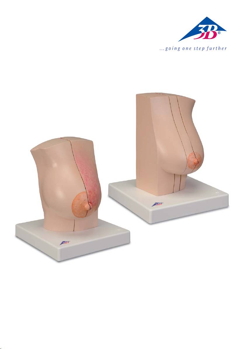
A Milk-giving right breast and chest wall, front view, medially divided, representation of the
externally visible changes for breast gland inflammation (mastitis) on the inner half
B Milk-giving right breast and chest wall, external half, cut surface
C Milk-giving right breast and chest wall, inner half, cut surface, representation of breast gland
inflammation (mastitis)
D Non-milk giving left breast and chest wall, front view, two sagittal cuts
E Non-milk-giving breast and chest wall; external half, external view,
skin windowed to illustrate the regional lymph nodes
F Non-milk-giving left breast and chest wall, external half, cut surface
G Non-milk-giving left breast and chest wall, middle section, external cut surface, representation
of a benign tumour (fibroadenoma) and two cysts
H Non-milk-giving left breast and chest wall, middle section, inner cut surface,
representation of a pathological proliferation of the breast gland connective tissue (mastopathy)
I Non-milk-giving left breast and chest wall, inner section, cut surface, representation of two
malignant tumours (breast cancer)
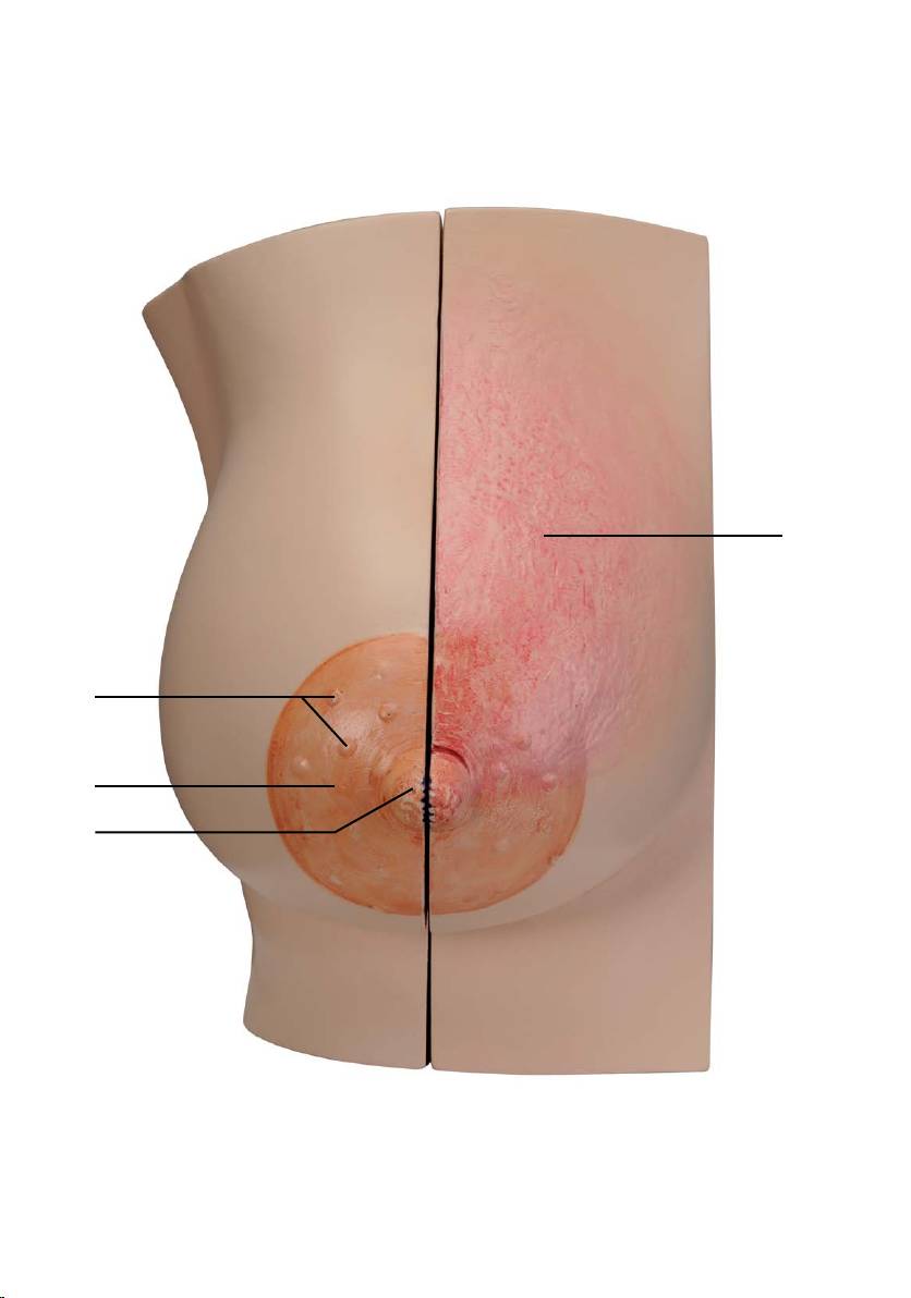
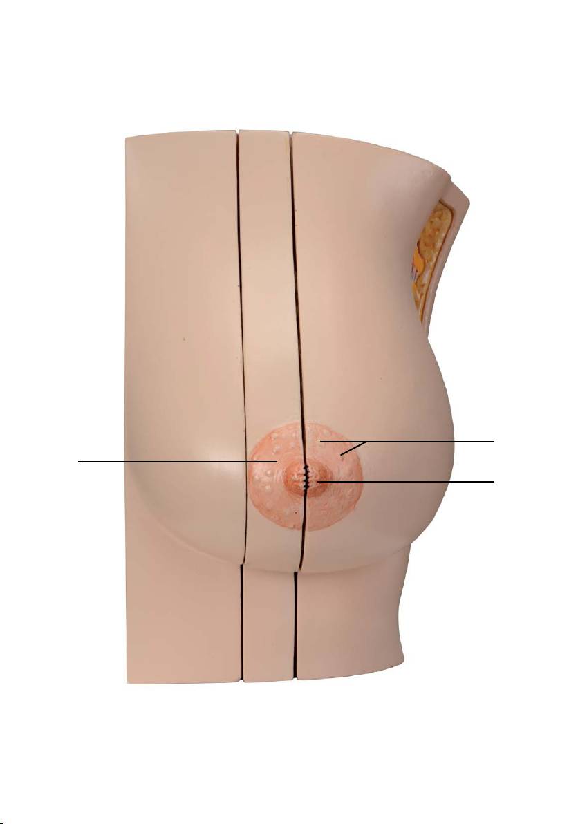
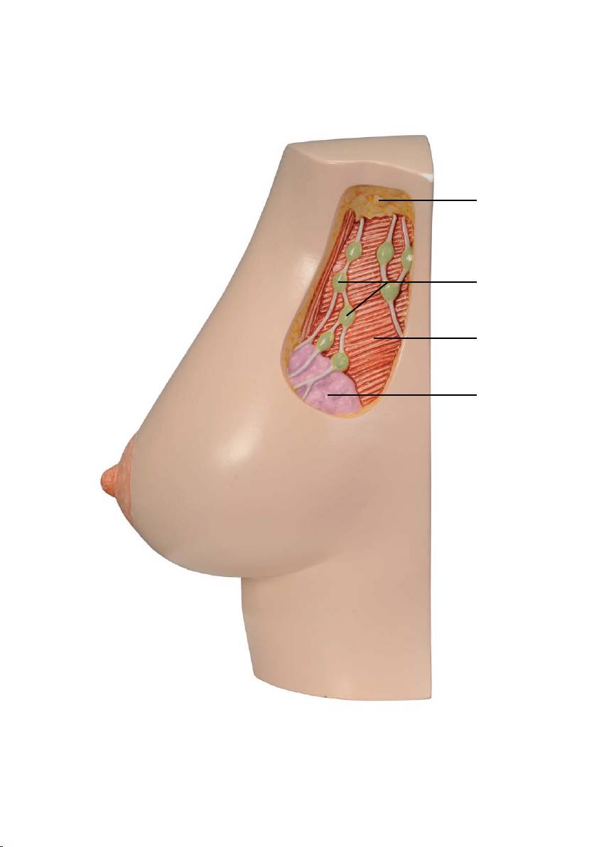
1 Tubercula areolae
2 Areola mammae
3 Papilla mammaria
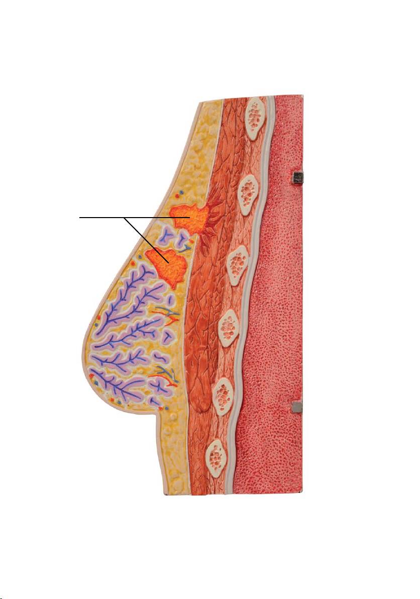
4 External signs of breast gland inflammation (Mastitis)
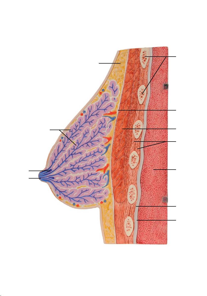
5 Fat tissue
6 Glandulae mammariae
7 Sinus lactiferi
8 Ductus lactiferi
9 Costae
10 Fascia pectoralis
11 M. pectoralis major
12 Mm. intercostales
13 Pulmo dexter
14 Pleura parietalis, Pars costalis
15 Pleura visceralis (Pleura pulmonalis)
16 Inflammatio (Mastitis)
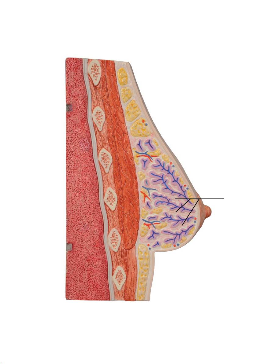
17 Nodi lymphoidei axillares, Nodi pectorales (Nodi anteriores)
18 Mamma
19 Ligamenta suspensoria mammaria (Retinaculum cutis mammae)
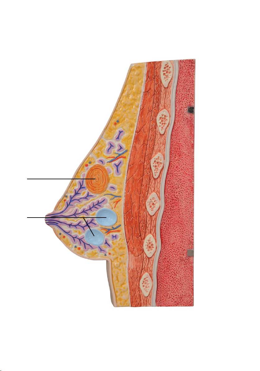
20 Pulmo sinister
21 Benign tumor (fibroadenoma)
22 Cystes
23 Proliferation of connective tissue (mastopathy)
24 Malignant tumors (breast cancer)
3

®
Model of female breast
English
L56 female breast and chest wall: milk-giving right breast with representation of an inflammation
(mastitis) and non-milk-giving left breast with representation of various diseases
The model consists of a milk-giving female right breast with the surrounding chest wall area and a non-
milk-giving female left breast with the surrounding chest wall area. Both parts of the model have a sagittal
cut. The cut surfaces show the tissue of the breast gland as well as the deeper-lying anatomical structures
such as the muscles, ribs, costal pleura, pulmonary pleura and lungs. A breast gland inflammation (masti-
tis) is represented on the right breast and various other diseases on the left breast.
The right breast and chest wall are separated into two halves by a sagittal cut in the mammary line and
these are held together by magnets. The cut area of the right half shows healthy breast tissue while the cut
area of the left half shows the changes to the breast gland when inflamed (mastitis). The exterior of this
half shows the associated pathological changes that are visible from the exterior.
The left breast and chest wall are divided into three parts by a sagittal cut in the mammary line and an
additional internal sagittal cut next to the areola and these are held together by magnets.
At the external part of the breast, the skin of the breast is windowed to view the regional lymph nodes. The
cut surface of this half show the healthy anatomical structures. The cut surfaces of the middle breast cut
show the various diseases of the breast gland. A benign tumour (fibroadenoma) and 2 cysts are represented
on the external cut surface. The inner cut surface shows a pathological proliferation of the breast connec-
tive tissue (mastopathy).
Two malignant tumours are represented in the breast gland on the cut surface of the inner part of the bre-
ast. One of these has extended into the chest wall.
L56 A
Milk-giving female right breast with representation of inflammation (mastitis)
The model shows a female right breast and the surrounding chest wall area. A sagittal cut in the mammary
line divides the model into two halves. These are held together with magnets. The cut surfaces of both hal-
ves of the model show the tissue of the breast gland as well as the deeper-lying anatomical structures such
as the muscles, ribs, costal pleura, pulmonary pleura and lungs. The cut area of the right half shows heal-
thy breast tissue while the cut area of the left half shows the changes to the breast gland when inflamed
(mastitis). The exterior of these two halves of the model shows the associated pathological changes that are
visible from the exterior.
L56 B
Non-milk-giving female left breast with representation of various diseases
The model shows a non-milk-giving female left breast and the surrounding chest wall area. A sagittal cut
in the mammary line and an additional internal sagittal cut next to the areola divides the model into
three parts. These are held together with magnets. All cut surfaces of the model parts show the tissue of
the breast gland as well as the deeper-lying anatomical structures such as the muscles, ribs, costal pleura,
pulmonary pleura and lungs.
At the outer part of the model, the skin of the breast is windowed to view the regional lymph nodes. The
cut surface of this half of the breast shows the healthy anatomical structures.
The cut surfaces of the middle cut of the breast show the various diseases of the breast gland. A benign
tumour (fibroadenoma) and 2 cysts are represented on the external cut surface. The inner cut surface
shows a pathological proliferation of the breast connective tissue (mastopathy).
Two malignant tumours are shown in the breast gland on the cut surface of the inner part of the model.
One of these has extended into the chest wall.
4

®
English
Model of female breast
A Milk-giving right breast and chest wall, front view, medially divided, representation of the
externally visible changes for breast gland inflammation (mastitis) on the inner half
B Milk-giving right breast and chest wall, external half, cut surface
C Milk-giving right breast and chest wall, inner half, cut surface, representation of breast gland
inflammation (mastitis)
D Non-milk giving left breast and chest wall, front view, two sagittal cuts
E Non-milk-giving breast and chest wall; external half, external view,
skin windowed to illustrate the regional lymph nodes
F Non-milk-giving left breast and chest wall, external half, cut surface
G Non-milk-giving left breast and chest wall, middle section, external cut surface, representation
of a benign tumour (fibroadenoma) and two cysts
H Non-milk-giving left breast and chest wall, middle section, inner cut surface,
representation of a pathological proliferation of the breast gland connective tissue (mastopathy)
I Non-milk-giving left breast and chest wall, inner section, cut surface, representation of two
malignant tumours (breast cancer)
1 Areolar tubercles (MONTGOMERY’s tubercles)
2 Areola
3 Nipple
4 External signs of breast gland inflammation (mastitis)
5 Fat tissue
6 Mammary glands
7 Lactiferous sinus
8 Lactiferous duct
9 Ribs
10 Pectoral fascia
11 Pectoralis major
12 Intercostal muscles
13 Right lung
14 Parietal pleura, costal part
15 Visceral pleura (Pulmonary pleura)
16 Inflammation (mastitis)
17 Axillary lymph nodes, pectoral nodes (anterior nodes)
18 Breast
19 Suspensory ligaments of breast (Suspensory retinaculum of breast)
20 Left lung
21 Benign tumor (fibroadenoma)
22 Cysts
23 Proliferation of connective tissue (mastopathy)
24 Malignant tumors (breast cancer)
5

®
Weibliches Brustmodell
Deutsch
L56 Weibliche Brust und Brustwand: milchgebende rechte Brust mit Darstellung einer Entzündung
(Mastitis), ruhende linke Brust mit Darstellung verschiedener Erkrankungen
Das Modell besteht aus einer rechten milchgebenden weiblichen Brust mit dem umgebenden
Brustwandbereich sowie einer linken ruhenden weiblichen Brust mit dem umgebenden Brustwandbereich.
Beide Modellteile sind sagittal geschnitten. Die Schnittflächen zeigen neben dem Gewebe der Brustdrüse
auch tiefer liegende anatomische Strukturen wie Muskeln, Rippen, Rippen- und Lungenfell und Lunge. An
der rechten Brust ist eine Brustdrüsenentzündung (Mastitis) dargestellt, die linke Brust zeigt verschiedene
Erkrankungen.
Durch einen Sagittalschnitt in der Mamillarlinie lassen sich rechte Brust und Brustwand in zwei Hälften
zerlegen, die durch Magnete zusammengehalten werden. Während die Schnittfläche der rechten Hälfte
gesundes Brustdrüsengewebe zeigt, sind auf der Schnittfläche der linken Hälfte die Veränderungen an der
Brustdrüse bei Entzündung (Mastitis) dargestellt. Die Außenseite dieser Hälfte zeigt die dazugehörenden
außen sichtbaren krankhaften Veränderungen.
Durch einen Sagittalschnitt in der Mamillarlinie und einen weiteren Sagittalschnitt innen neben dem
Brustwarzenvorhof lassen sich linke Brust und Brustwand in drei Teile zerlegen, die durch Magnete
zusammengehalten werden.
An dem äußeren Brustteil ist die Brusthaut zur Ansicht der regionären Lymphknoten gefenstert. Die
Schnittfläche dieser Hälfte zeigt die gesunden anatomischen Strukturen. Die Schnittflächen des mittleren
Brustausschnitts zeigen verschiedene Erkrankungen der Brustdrüse. Auf der äußeren Schnittfläche sind
neben einem gutartigen Tumor (Fibroadenom) 2 Zysten dargestellt. Die innere Schnittfläche zeigt eine
krankhafte Vermehrung des Brustbindegewebes (Mastopathie).
Auf der Schnittfläche des inneren Brustteils sind zwei bösartige Tumoren in der Brustdrüse dargestellt,
von denen sich einer in die Brustwand ausgedehnt hat.
L56 A
Milchgebende rechte weibliche Brust mit Darstellung einer Entzündung (Mastitis)
Das Modell zeigt eine rechte weibliche Brust und den umgebenden Brustwandbereich. Durch einen
Sagittalschnitt in der Mamillarlinie besteht das Modell aus zwei Hälften. Diese werden durch Magnete
zusammengehalten. Auf den Schnittflächen beider Modellhälften sind neben dem Gewebe der Brustdrüse
auch die tiefer liegenden anatomischen Strukturen wie Muskeln, Rippen, Rippen- und Lungenfell und
Lunge dargestellt. Während die Schnittfläche der rechten Hälfte gesundes Brustdrüsengewebe zeigt, sind
auf der Schnittfläche der linken Hälfte die Veränderungen an der Brustdrüse bei Entzündung (Mastitis)
dargestellt. Die Außenseite dieser Modellhälfte zeigt die dazugehörenden außen sichtbaren krankhaften
Veränderungen.
L56 B
Ruhende linke weibliche Brust mit Darstellung verschiedener Erkrankungen
Das Modell zeigt eine ruhende linke weibliche Brust und den umgebenden Brustwandbereich. Durch einen
Sagittalschnitt in der Mamillarlinie und einen weiteren Sagittalschnitt innen neben dem Brustwarzenvorhof
besteht das Modell aus drei Teilen. Diese werden durch Magnete zusammengehalten. Alle Schnittflächen
der Modellteile zeigen neben dem Gewebe der Brustdrüse auch tiefer liegende anatomische Strukturen wie
Muskeln, Rippen, Rippen- und Lungenfell und Lunge.
An dem äußeren Modellteil ist die Brusthaut zur Ansicht der regionären Lymphknoten gefenstert. Die
Schnittfläche dieser Brusthälfte zeigt die gesunden anatomischen Strukturen.
Die Schnittflächen des mittleren Modellausschnitts zeigen verschiedene Erkrankungen der Brustdrüse.
Auf der äußeren Schnittfläche sind neben einem gutartigen Tumor (Fibroadenom) 2 Zysten dargestellt.
Die innere Schnittfläche zeigt eine krankhafte Vermehrung des Brustbindegewebes (Mastopathie).
Auf der Schnittfläche des inneren Modellteils sind zwei bösartige Tumoren in der Brustdrüse dargestellt,
von denen sich einer in die Brustwand ausgedehnt hat.
6

®
Deutsch
Weibliches Brustmodell
A Milchgebende rechte Brust und Brustwand, von vorne, median geschnitten, Darstellung
der außen sichtbaren Veränderungen bei Brustdrüsenentzündung (Mastitis) auf der inneren
Hälfte
B Milchgebende rechte Brust und Brustwand, äußere Hälfte, Schnittfläche
C Milchgebende rechte Brust und Brustwand, innere Hälfte, Schnittfläche, Darstellung einer
Brustdrüsenentzündung (Mastitis)
D Ruhende linke Brust und Brustwand, von vorne, zweifach sagittal geschnitten
E Ruhende linke Brust und Brustwand, äußere Hälfte, von außen, Haut zur Darstellung der
regionären Lymphknoten gefenstert
F Ruhende linke Brust und Brustwand, äußere Hälfte, Schnittfläche
G Ruhende linke Brust und Brustwand, mittlerer Abschnitt, äußere Schnittfläche,
Darstellung eines gutartigen Tumors (Fibroadenom) und zweier Zysten
H Ruhende linke Brust und Brustwand, mittlerer Abschnitt, innere Schnittfläche,
Darstellung einer krankhaften Vermehrung des Brustdrüsenbindegewebes (Mastopathie)
I Ruhende linke Brust und Brustwand, innerer Abschnitt, Schnittfläche, Darstellung zweier
bösartiger Tumoren (Brustkrebs)
1 Erhebungen der Warzenhofdrüsen (MONTGOMERY-Knötchen)
2 Warzenhof
3 Brustwarze
4 Äußere Zeichen einer Brustdrüsenentzündung (Mastitis)
5 Fettgewebe
6 Brustdrüsen
7 Aufweitung des Milchgangs (Milchsäckchen)
8 Milchgang
9 Rippen
10 Bindegewebige Umhüllung (Faszie) des großen Brustmuskels
11 Großer Brustmuskel
12 Zwischenrippenmuskeln
13 Rechte Lunge
14 Rippenfell
15 Lungenfell
16 Entzündung (Mastitis)
17 Achsellymphknoten, Lymphknoten auf dem großen Brustmuskel (Vordere Lymphknoten)
18 Brustdrüse
19 Brustdrüsenaufhängebänder
20 Linke Lunge
21 Gutartiger Tumor (Fibroadenom)
22 Zysten
23 Vermehrung des Bindegewebes (Mastopathie)
24 Bösartige Tumoren (Brustkrebs)
7

®
EspañolModelo de mama de mujer
L56 Mama de mujer y pared torácica: mama derecha lactante con representación de una inflamación
(mastitis), mama izquierda en reposo con representación de distintas enfermedades
El modelo se compone de una mama derecha lactante de mujer con región torácica circundante y una
mama izquierda de mujer en reposo con región torácica circundante. El corte de ambos modelos es sagital.
Las secciones de corte muestran, además del tejido de la glándula mamaria, estructuras anatómicas más
profundas, como músculos, costillas, pleura y pleura pulmonar y pulmón. En la mama derecha se repre-
senta una inflamación mamaria (mastitis) y la mama izquierda muestra distintas enfermedades.
El corte sagital por la línea mamilar permite desmontar la mama derecha y la pared torácica en dos mita-
des que se mantienen unidas por medio de imanes. La superficie de corte de la mitad derecha muestra
tejido glandular mamario sano y la sección de corte de la mitad izquierda reproduce los cambios en la
glándula mamaria en caso de inflamación (mastitis). El lado externo de esta mitad presenta los correspon-
dientes cambios patológicos visibles.
El corte sagital por la línea mamilar y un corte sagital adicional interno junto a la areola permiten desmon-
tar la mama izquierda y la pared torácica en tres piezas, que se mantienen unidas por medio de imanes.
La parte externa de la mama presenta una abertura en forma de ventana para poder ver los ganglios lin-
fáticos de esa región. La superficie de corte de esta mitad muestra las estructuras anatómicas sanas. Las
superficies de corte de la sección central de la mama presentan distintas enfermedades de la glándula
mamaria. En la superficie externa se representan 2 quistes y un tumor benigno (fibroadenoma). La superfi-
cie de corte interna muestra una proliferación patológica del tejido conectivo de la mama (mastopatía).
En la superficie de corte del interior de la mama hay dos tumores malignos en la glándula mamaria, de los
cuales uno se extiende hasta la pared torácica.
L56 A
Mama derecha lactante de mujer con representación de una inflamación (mastitis)
El modelo muestra la mama derecha de una mujer y la región de la pared torácica circundante. El modelo
consta de dos mitades divididas por un corte sagital en la línea mamilar. Las dos mitades se mantienen
unidas con imanes. En las superficies de corte de las dos mitades del modelo se muestran, además del
tejido de la glándula mamaria, estructuras anatómicas más profundas, como músculos, costillas, pleura y
pleura pulmonar y pulmón. La superficie de corte de la mitad derecha muestra tejido glandular mamario
sano y la sección de corte de la mitad izquierda reproduce los cambios en la glándula mamaria en caso de
inflamación (mastitis). El lado externo de esta mitad del modelo presenta los correspondientes cambios
patológicos visibles.
L56 B
Mama izquierda en reposo de mujer con representación de distintas enfermedades
El modelo muestra la mama izquierda en reposo de una mujer y la región de la pared torácica circundante.
El modelo se compone de tres piezas divididas por un corte sagital por la línea mamilar y un corte sagital
complementario en el interior junto a la areola. Las piezas se mantienen unidas con imanes. Todas las sec-
ciones de corte de las piezas del modelo muestran, además del tejido de la glándula mamaria, estructuras
anatómicas más profundas, como músculos, costillas, pleura y pleura pulmonar y pulmón.
La parte externa del modelo presenta una abertura en forma de ventana para poder ver los ganglios lin-
fáticos de esa región. La superficie de corte de esta mitad de la mama muestra las estructuras anatómicas
sanas.
Las superficies de corte de la sección central del modelo presentan distintas enfermedades de la glándula
mamaria. En la superficie externa se representan 2 quistes y un tumor benigno (fibroadenoma). La superfi-
cie de corte interna muestra una proliferación patológica del tejido conectivo de la mama (mastopatía).
En la superficie de corte del interior del modelo hay dos tumores malignos en la glándula mamaria, de los
cuales uno se extiende hasta la pared torácica.
8

®
Español Modelo de mama de mujer
A Mama lactante derecha y pared torácica, vista frontal, corte medial, representación de los
cambios externos visibles con inflamación de glándula mamaria (mastitis) en la mitad interna
B Mama lactante derecha y pared torácica, mitad externa, superficie de corte
C Mama lactante derecha y pared torácica, mitad interna, superficie de corte,
representación de una inflamación de la glándula mamaria (mastitis)
D Mama izquierda en reposo y pared torácica, vista frontal, doble corte sagital
E Mama izquierda en reposo y pared torácica; mitad externa, vista externa,
piel con ventana para la visualización de los ganglios linfáticos de la región
F Mama izquierda en reposo y pared torácica, mitad externa, superficie de corte
G Mama izquierda en reposo y pared torácica, sección medial, superficie de corte externa,
representación de un tumor benigno (fibroadenoma) y dos quistes
H Mama izquierda en reposo y pared torácica, sección medial, superficie de corte interna,
representación de una proliferación patológica del tejido conectivo de la glándula mamaria
(mastopatía)
I Mama izquierda en reposo y pared torácica, sección interna, superficie de corte,
representación de dos tumores malignos (cáncer de mama)
1 Eminencias en las glándulas de la aureola (Glándulas de MONTGOMERY)
2 Areola
3 Pezón
4 Signos externos de inflamación de la glándula mamaria (mastitis)
5 Tejido adiposo
6 Glándulas mamarias
7 Dilatación del conducto galactóforo (seno lactífero)
8 Conducto galactóforo
9 Costillas
10 Envoltura de tejido conectivo (fascia) del músculo pectoral mayor
11 Músculo pectoral mayor
12 Músculos intercostales
13 Pulmón derecho
14 Pleura
15 Pleura pulmonar
16 Inflamación (mastitis)
17 Ganglio linfático axilar, ganglio linfático en el músculo pectoral mayor (ganglio linfático anterior)
18 Glándula mamaria
19 Ligamentos suspensorios de la glándula mamaria
20 Pulmón izquierdo
21 Tumor benigno (fibroadenoma)
22 Quistes
23 Proliferación de tejido conectivo (mastopatía)
24 Tumores malignos (cáncer de mama)
9

®
FrançaisModèle de poitrine féminine
Poitrine féminine et paroi thoracique L56 : poitrine droite donnant le sein avec représentation d’une
inflammation (Mastitis), poitrine gauche au repos avec représentation de diverses affections
Le modèle anatomique est constitué d’une poitrine droite féminine donnant le sein comprenant
l’environnement de la paroi thoracique ainsi que d’une poitrine gauche au repos et de son environnement
de la paroi thoracique. Les deux parties du modèle anatomique sont représentées en coupe Les surfaces
de coupe montrent de profondes structures anatomiques parallèlement au tissu de la glande mammaire,
ainsi que les muscles, les côtes, la plèvre des côtes et la plèvre pulmonaire et les poumons. Une inflamma-
tion des glandes mammaires de la poitrine droite est également représentée (mastitis), la poitrine gauche
met en évidence diverses affections. Un plan sagittal de la ligne mammaire sépare la poitrine droite et la
paroi thoracique en deux moitiés qui sont maintenues ensemble par un système d’aimantation. Alors que
le plan sagittal droit met en évidence un tissu de glandes mammaires sain, le plan sagittal gauche repré-
sente les modifications de la glande mammaire suite à une inflammation (mastitis). La face externe de
cette moitié met en évidence les modifications externes notoires s’y rapportant.
Un plan sagittal de la ligne mammaire et un plan sagittal interne supplémentaire parallèlement au mame-
lon mammaire mettent en évidence la poitrine gauche et la paroi thoracique en trois parties, maintenues
ensemble par un système d’aimantation. La peau de la poitrine de la partie externe du sein est représentée
dans une fenêtre afin de pouvoir visualiser les noeuds lymphatiques de cette zone. La surface de coupe
de cette moitié met en évidence les structures anatomiques saines. Les surfaces de la coupe moyenne de
la poitrine mettent en évidence diverses affections de la glande mammaire. 2 kystes sont représentés sur
la surface de coupe externe, parallèlement à une tumeur bénine (adénome fibreux). La surface de coupe
interne met en évidence une multiplication de la dégénérescence maladive du tissu conjonctif (mastopa-
thie). Deux tumeurs malignes de la glande mammaire sont représentées sur la surface de coupe de la par-
tie interne du sein, dont une s’est étendue à la paroi thoracique.
L56 A
Poitrine droite donnant le sein avec représentation d’une inflammation (mastitis)
Le modèle anatomique met en évidence une poitrine droite féminine et l’environnement de la paroi
thoracique. Grâce à un plan sagittal de la ligne mammaire, le modèle anatomique est constitué de deux
moitiés. Ces dernières sont maintenues ensemble par un système d’aimantation. Les surfaces de coupe des
deux moitiés du modèle anatomique mettent également en évidence de profondes structures anatomiques
parallèlement au tissu de la glande mammaire, ainsi que des muscles, les côtes, la plèvre des côtes et la
plèvre pulmonaire et les poumons. Alors que le plan sagittal droit met en évidence un tissu de glandes
mammaires sain, le plan sagittal gauche représente les modifications de la glande mammaire suite à une
inflammation (mastitis). La face externe de cette moitié de modèle anatomique met en évidence les modi-
fications externes notoires s’y rapportant.
L56 B
Poitrine gauche féminine au repos avec représentation de diverses affections
Le modèle anatomique met en évidence une poitrine gauche féminine et l’environnement de la paroi tho-
racique. Un plan sagittal de la ligne mammaire et un plan sagittal interne supplémentaire parallèlement
au mamelon donnent au modèle anatomique trois parties. Ces dernières sont maintenues ensemble par un
système d’aimantation. Les surfaces de coupe montrent également de profondes structures anatomiques
parallèlement au tissu de la glande mammaire, ainsi que des muscles, les côtes, la plèvre des côtes et la
plèvre pulmonaire et les poumons. La peau de la poitrine de la partie externe du sein est représentée dans
une fenêtre afin de pouvoir visualiser les noeuds lymphatiques de cette zone. La surface de coupe de cette
moitié de poitrine met en évidence les structures anatomiques saines. Les surfaces de la coupe moyenne de
la poitrine mettent en évidence diverses affections de la glande mammaire. 2 kystes sont représentés sur
la surface de coupe externe, parallèlement à une tumeur bénine (adénome fibreux). La surface de coupe
interne met en évidence une multiplication de la dégénérescence maladive du tissu conjonctif (mastopa-
thie). Deux tumeurs malignes de la glande mammaire sont représentées sur la surface de coupe de la par-
tie interne du sein, dont une s’est étendue à la paroi thoracique.
10

®
Français Modèle de poitrine féminine
A Coupe avant médiane de poitrine droite donnant le sein et paroi thoracique, représentation des
modifications externes notoires en cas d’inflammation des glandes mammaires (mastitis) de la
face interne
B Poitrine droite donnant le sein et paroi thoracique, moitié externe, surface de coupe
C Poitrine droite donnant le sein et paroi thoracique, moitié interne, surface de coupe,
représentation d’une inflammation des glandes mammaires (mastitis)
D Poitrine gauche au repos et paroi thoracique, plan sagittal double, frontal
E Poitrine gauche au repos et paroi thoracique ; moité externe, vue externe, peau pour
représentation des noeuds lymphatiques de la zone, visualisation dans une fenêtre
F Poitrine gauche donnant le sein et paroi thoracique, moitié externe, surface de coupe
G Poitrine gauche au repos et paroi thoracique, partie moyenne, surface de coupe externe,
représentation d’une tumeur bénine (adénome fibreux) et de deux kystes
H Poitrine gauche au repos et paroi thoracique, partie moyenne, surface de coupe interne,
représentation d’une multiplication de la dégénérescence maladive du tissu conjonctif des
glandes mammaires (mastopathie)
I Poitrine gauche au repos et paroi thoracique, partie interne, surface de coupe,
représentation de deux tumeurs malignes (cancer du sein)
1 Soulèvements des glandes des mamelons (nodules de MONTGOMERY)
2 Mamelon
3 Aréole du sein
4 Signes extérieurs d’une inflammation des glandes mammaires (mastitis)
5 Tissu adipeux
6 Glandes mammaires
7 Expansion du canal galactophore (Acini)
8 Canal galactophore
9 Côtes
10 Enveloppe du tissu conjonctif (fascia) du muscle mammaire principal
11 Grand pectoral
12 Muscles intercostaux
13 Poumon droit
14 Plèvre des côtes
15 Plèvre pulmonaire
16 Inflammation (mastitis)
17 Noeuds lymphatiques de l’aisselle, noeuds lymphatiques situés sur le muscle mammaire principal
(noeuds lymphatiques frontaux)
18 Glande mammaire
19 Ligaments de Cooper des glandes mammaires
20 Poumon gauche
21 Tumeur bénine (adénome fibreux)
22 Kystes
23 Multiplication du tissu conjonctif (mastopathie)
24 Tumeurs malignes (cancer du sein)
11
A
4
1
2
3
12
B
9
5
10
11
6
12
13
7
8
14
15
13

C
16
14
D
1
2
3
15
E
5
17
11
18
16
F
9
11
5
19
12
6
20
7
8
14
15
17
G
21
22
18
H
23
19
I
24
20

®
Português Modelo de Mama Feminina
L56 Mama feminina e parede torácica: mama direita lactante com representação de inflamação
(mastite), mama esquerda não lactante com representação de diversas doenças
O modelo é composto de mama feminina direita lactante com a área da parede torácica adjacente, bem
como de mama feminina esquerda não lactante com a área da parede torácica adjacente. Ambos os mode-
los são seccionados sagitalmente. Além dos tecidos da glândula mamária, as seções mostram também
estruturas anatômicas mais profundas, como músculos, costelas, pleura parietal, pleura visceral e pulmões.
A mama direita ilustra inflamação de glândula mamária (mastite), ao passo que a mama esquerda mostra
diversas doenças.
Através de seção sagital pela linha mamária, pode-se desmontar a mama direita e a parede torácica em
duas metades, que são mantidas unidas por ímãs. Enquanto a seção da metade direita mostra tecido
mamário saudável, a seção da metade esquerda mostra as alterações na glândula mamária devido à
inflamação (mastite). O lado externo desta metade mostra as alterações patológicas pertinentes visíveis
externamente.
Através da seção sagital pela linha mamária e uma seção sagital adicional interna ao lado da aréola, pode-
se desmontar a mama esquerda e a parede torácica em três partes, que são mantidas unidas por ímãs.
Do lado externo da mama, a pele foi fenestrada ara permitir a visualização dos linfonodos regionais. A
seção desta metade ilustra as estruturas anatômicas sadias. As áreas de corte do segmento mamário medial
ilustram diversas patologias da glândula mamária. Na seção externa, além de ilustrar um tumor benigno
(fibroadenoma), também mostra 2 cistos.
A seção interna mostra a multiplicação patológica do tecido conjuntivo mamário (mastopatia).
Sobre a seção do segmento mamário interno são representados dois tumores malignos na glândula
mamária, um dos quais se espalhou para a parede torácica.
L56 A
Mama feminina direita lactante com representação de inflamação (mastite)
O modelo mostra uma mama feminina direita e a região da parede torácica adjacente. Através de corte
sagital pela linha mamária, o modelo se divide em duas metades. Estas metades são mantidas unidas por
ímãs. As áreas de corte de ambas as metades mostram além do tecido da glândula mamária também as
estruturas anatômicas mais profundas, como músculos, costelas, pleura parietal, pleura visceral e pulmões.
Enquanto a seção da metade direita mostra tecido mamário saudável, a seção da metade esquerda mostra
as alterações na glândula mamária devido à inflamação (mastite). O lado externo destas metades mostra as
alterações patológicas pertinentes visíveis externamente.
L56 B
Mama feminina esquerda não lactante com representação de diversas patologias
O modelo mostra uma mama feminina esquerda não lactante e a parede torácica adjacente. Através da
seção sagital pela linha mamária e uma seção sagital adicional interna ao lado da aréola, o modelo se divi-
de em três partes. Estas partes são mantidas unidas por ímãs. Todas as áreas de corte das partes do modelo
mostram além dos tecidos da glândula mamária também estruturas anatômicas mais profundas, como
músculos, costelas, pleura parietal, pleura visceral e pulmões.
No segmento externo da mama, a pele foi fenestrada para permitir a visualização dos linfonodos regionais.
A seção desta parte ilustra as estruturas anatômicas sadias.
As áreas de corte do segmento medial do modelo ilustram diversas patologias da glândula mamária. Na
seção externa, além de ilustrar um tumor benigno (fibroadenoma), também mostra 2 cistos. A seção inter-
na mostra a multiplicação patológica do tecido conjuntivo mamário (mastopatia).
Sobre a seção do segmento mamário interno são representados dois tumores malignos na glândula
mamária, um dos quais se espalhou para a parede torácica.
21

®
PortuguêsModelo de Mama Feminina
A Mama direita lactante e parede torácica, vista frontal, com seção mediana, representação das
alterações visíveis externamente durante inflamação da glândula mamária (mastite) no segmen-
to interno
B Mama direita lactante e parede torácica, metade externa, seção
C Mama direita lactante e parede torácica, metade interna, seção, representação de inflamação
da glândula mamária (mastite)
D Mama esquerda não lactante e parede torácica, vista anterior, secção sagital dupla
E Mama esquerda não lactante e parede torácica, vista frontal, seccionada sagitalmente duas
vezes
F Mama esquerda não lactante e parede torácica; metade externa, vista externa, pele foi fenestra-
da para ilustrar os linfonodos regionais
G Mama esquerda não lactante e parede torácica, metade externa, seção
H Mama esquerda não lactante e parede torácica, segmento medial, seção externa, representação
de tumor benigno (fibroadenoma) e dois cistos
I Mama esquerda não lactante e parede torácica, segmento medial, seção interna, representação
da multiplicação patológica do tecido conjuntivo da glândula mamária (mastopatia)
1 Saliências das glândulas das aréolas (tubérculos de MONTGOMERY)
2 Aréola
3 Mamilo
4 Sinais externos de inflamação da glândula mamária (mastite)
5 Tecido adiposo
6 Glândulas mamárias
7 Dilatação do duto lactífero (seios lactíferos)
8 Duto lactífero
9 Costelas
10 Revestimento de tecido conjuntivo (fáscia) do músculo peitoral maior
11 Músculo peitoral maior
12 Músculos intercostais
13 Pulmão direito
14 Pleura parietal
15 Pleura visceral
16 Inflamação (mastite)
17 Linfonodos axilares, linfonodos sobre o músculo peitoral maior (linfonodos anteriores)
18 Glândula mamária
19 Ligamentos suspensores da glândula mamária
20 Pulmão esquerdo
21 Tumor benigno (fibroadenoma)
22 Cistos
23 Multiplicação do tecido conjuntivo (mastopatia)
24 Tumores malignos (câncer de mama)
22

®
Italiano Modello di torso femminile
L56 Torso e parete toracica femminile: seno destro lattifero con rappresentazione di
un’infiammazione (mastite), seno sinistro statico con rappresentazione di diverse patologie
Il modello è costituito da un seno destro lattifero femminile e da un seno sinistro statico, entrambi con
la parete toracica circostante. Le due parti del modello sono sezionate sagittalmente. Le superfici delle
sezioni mostrano, accanto al tessuto della ghiandola mammaria, strutture anatomiche più profonde
come i muscoli, le costole, la pleura parietale e polmonare e il polmone. Sul seno destro è rappresentata
un’infiammazione della ghiandola mammaria (mastite), il seno sinistro mostra diverse patologie.
Attraverso una sezione sagittale nella linea mammaria è possibile scomporre il seno destro e la parete
toracica in due parti, che vengono unite tramite magneti. Mentre la superficie della metà destra mostra un
tessuto della ghiandola mammaria sano, sulla superficie della metà sinistra sono mostrate le alterazioni
sulla ghiandola dovute all’infiammazione (mastite). La parte esterna di questa metà mostra le relative
alterazioni patologiche esterne visibili.
Attraverso una sezione sagittale nella linea mammaria e un’ulteriore sezione sagittale interna accanto
all’areola è possibile scomporre il seno sinistro e la parete toracica in tre parti, che vengono tenute unite
tramite magneti.
Sulla parte esterna del seno, la pelle presenta un’apertura che consente di osservare i linfonodi regionali.
La superficie di questa metà mostra le strutture anatomiche sane. Le superfici della sezione centrale del
seno mostrano varie patologie della ghiandola mammaria. Sulla superficie esterna sono rappresentati un
tumore benigno (fibroadenoma) e 2 cisti. La superficie interna mostra un ingrossamento patologico del
tessuto connettivo della mammella (mastopatia).
Sulla superficie interna del seno sono rappresentati due tumori maligni nella ghiandola mammaria, uno
dei quali si è esteso alla parete toracica.
L56 A
Seno destro lattifero femminile con rappresentazione di un’infiammazione (mastite)
Il modello mostra un seno destro femminile e la parete toracica circostante. Attraverso una sezione sagit-
tale nella linea mammaria, il modello è costituito da due metà che vengono unite da magneti. Sulle super-
fici delle metà sono rappresentate, accanto al tessuto della ghiandola mammaria, strutture anatomiche
più profonde come i muscoli, le costole, la pleura parietale e polmonare e il polmone. Mentre la superficie
della metà destra mostra un tessuto della ghiandola mammaria sano, sulla superficie della metà sinistra
sono mostrate le alterazioni sulla ghiandola dovute all’infiammazione (mastite). La parte esterna di questa
metà mostra le relative alterazioni patologiche esterne visibili.
L56 B
Seno femminile sinistro statico con rappresentazione di diverse patologie
Il modello mostra un seno sinistro femminile statico e la parete toracica circostante. Attraverso una sezio-
ne sagittale nella linea mammaria e un’ulteriore sezione sagittale interna accanto all’areola, il modello è
costituito da tre parti, che vengono unite da magneti. Tutte le superfici delle parti del modello mostrano,
accanto al tessuto della ghiandola mammaria, strutture anatomiche più profonde come i muscoli, le costo-
le, la pleura parietale e polmonare e il polmone.
Sulla parte esterna del modello, la pelle presenta un’apertura che consente di osservare i linfonodi regio-
nali. La superficie di questa metà del seno mostra le strutture anatomiche sane.
Le superfici della sezione centrale del modello mostrano varie patologie della ghiandola mammaria. Sulla
superficie esterna sono rappresentati un tumore benigno (fibroadenoma) e 2 cisti. La superficie interna
mostra un ingrossamento patologico del tessuto connettivo della mammella (mastopatia).
Sulla superficie interna del modello sono rappresentati due tumori maligni nella ghiandola mammaria,
uno dei quali si è esteso alla parete toracica.
23

®
ItalianoModello di torso femminile
A Seno destro lattifero e parete toracica, vista anteriore, sezione mediana, rappresentazione delle
alterazioni patologiche esterne visibili in caso di infiammazione della ghiandola mammaria
(mastite) sulla metà interna
B Seno destro lattifero e parete toracica, metà esterna, superficie della sezione
C Seno destro lattifero e parete toracica, metà interna, superficie della sezione,
rappresentazione di un’infiammazione della ghiandola mammaria (mastite)
D Seno sinistro statico e parete toracica, vista anteriore, sezione sagittale doppia
E Seno sinistro statico e parete toracica, metà esterna, vista esterna, pelle con apertura che
consente di osservare i linfonodi regionali
F Seno sinistro statico e parete toracica, metà esterna, superficie della sezione
G Seno sinistro statico e parete toracica, sezione centrale, superficie esterna della sezione,
rappresentazione di un tumore benigno (fibroadenoma) e due cisti
H Seno sinistro statico e parete toracica, sezione centrale, superficie interna della sezione,
rappresentazione di un ingrossamento patologico del tessuto connettivo della mammella
(mastopatia)
I Seno sinistro statico e parete toracica, sezione interna, superficie della sezione,
rappresentazione di due tumori maligni (cancro al seno)
1 Ingrossamento delle ghiandole dell’areola (tubercoli di Montgomery)
2 Areola
3 Capezzolo
4 Segni esterni di un’infiammazione della ghiandola mammaria (mastite)
5 Tessuto adiposo
6 Ghiandole mammarie
7 Dilazione del dotto glattoforo (pori lattiferi)
8 Dotto glattoforo
9 Costole
10 Fascia del tessuto connettivo del muscolo pettorale maggiore
11 Muscolo pettorale maggiore
12 Muscoli intercostali
13 Polmone destro
14 Pleura parietale
15 Pleura polmonare
16 Infiammazione (mastite)
17 Linfonodi ascellari, linfonodi sul muscolo pettorale maggiore (linfonodi anteriori)
18 Ghiandola mammaria
19 Legamento pettineo
20 Polmone sinistro
21 Tumore benigno (fibroadenoma)
22 Cisti
23 Ingrossamento del tessuto connettivo (mastopatia)
24 Tumori maligni (cancro al seno)
24

日本語
乳房疾患モデル(授乳期・非授乳期)
L56 女性乳房と胸壁:乳腺炎を表示する右乳房(授乳期)と様々な症状を表示する左乳房(非授乳期)
このモデルは授乳期の右乳房と非授乳期の左乳房を再現しており,両方とも矢状断で分割できます。
断面は乳房組織の他に筋肉,肋骨,肋骨胸膜,肺胸膜,肺など解剖学構造も表示しています。
右乳房では乳腺炎,左乳房ではその他の様々な症状を確認できます。
右乳房は中央から矢状断で分割された2つのパーツからなり,それぞれは磁石で固定されています。断面右側
では健康な授乳期の乳房組織,断面左側では乳腺炎が確認できます。また表面では病理学的変化を見ることが
できます。
左乳房は2箇所の矢状断で3つに分解でき,こちらもそれぞれを磁石で固定しています。
外側パーツ:皮膚の一部は開放されており,所属リンパ節を見ることができます。断面は健康な乳房の解剖学
的構造を再現しています。
中央パーツ:断面では様々な症状が確認でき,外側断面には良性腫瘍(線維腺腫)と嚢胞,内側断面には乳房
結合組織の進行病変(乳腺繊維嚢胞症)を表示しています。
内側パーツ:断面では2つの悪性腫瘍を表示しており,そのうち1つは胸壁に達しています。
右乳房
乳腺炎をを表示した,授乳期の右乳房
乳腺炎をもつ女性の右乳房を再現。
矢状断で中央から2つに分解でき,それぞれは磁石で固定されています。両側の断面には,乳房組織の他に筋
肉,肋骨,肋骨胸膜,肺胸膜,肺など解剖学構造を表示しています。
断面右側では健康な乳房組織を表示,左側では乳腺炎を表示しており,皮膚で病理学的変化も見ることができ
ます。
左乳房
様々な症状を表示する,非授乳期の左乳房
非授乳期の左乳房組織を再現。
2箇所の矢状断で3つに分解でき,それぞれは磁石で固定されています。
すべての断面で乳房組織の他に筋肉,肋骨,肋骨胸膜,肺胸膜,肺など解剖学構造を表示しています。
外側パーツ:皮膚の一部は開放されており,所属リンパ節を確認できます。断面には健康な乳房の解剖学的構
造を表示。
中央パーツ:断面で様々な症状を確認できます。外側断面に良性腫瘍(線維腺腫)と嚢胞を表示,内側断面に
は乳房結合組織の進行病変(乳腺繊維嚢胞症)を表示してます。
内側パーツ:断面には2つの悪性腫瘍を表示。そのうち1つは胸壁に達しています。
25

乳房疾患モデル(授乳期・非授乳期)
日本語
A 授乳期の右乳房,前面,中央で分割。内側半分には外観から認識できる乳腺炎を表示
B 授乳期の右乳房,外側半分の断面
C 授乳期の右乳房,内側半分の断面,乳腺炎を表示
D 非授乳期の左乳房,前面,2箇所で分割
E 非授乳期の左乳房,外側パーツの概観,皮膚の開放部で所属リンパ節を表示
F 非授乳期の左乳房,外側パーツの断面
G 非授乳期の左乳房,中央パーツの外側断面,良性腫瘍(線維腺腫)と嚢胞を表示
H 非授乳期の左乳房,中央パーツの内側断面,乳房結合組織の進行病変(乳腺繊維嚢胞症)を表示
I 非授乳期の左乳房,内側パーツの断面。悪性腫瘍(乳癌)を表示
1 乳輪結節(モンゴメリー結節)
2 乳輪
3 乳頭
4 乳腺炎の外部兆候
5 脂肪組織
6 乳腺
7 乳管洞
8 乳管
9 肋骨
10 胸筋筋膜
11 大胸筋
12 肋間筋
13 右肺
14 壁側胸膜
15 臓側胸膜 (肺胸膜)
16 乳腺炎
17 腋窩リンパ節
18 乳房
19 乳房提靱帯
20 左肺
21 良性腫瘍(線維腺腫)
22 嚢胞
23 乳房結合組織の進行病変(乳腺繊維嚢胞症)
24 悪性腫瘍(乳癌)
26

®
Русский Модель женской молочной железы
L56 – женская грудная стенка и молочная железа: правая молочная железа в состоянии
лактации с признаками воспаления (мастит) и левая молочная железа вне лактации с
признаками различных заболеваний
Модель состоит из правой женской молочной железы в состоянии лактации и окружающей ее частью
грудной стенки и левой женской молочной железы вне лактации и окружающей ее частью грудной
стенки. Обе части модели представлены в сагиттальном разрезе. Поверхности среза представлены
тканью молочной железы, а также лежащими глубже анатомическими структурами, такими как мышцы,
ребра, костальная и висцеральная плевра и легкие. Воспаление молочной железы (мастит) представлено
на правой молочной железе, а различные другие заболевания – на левой. Правая молочная железа
и грудная стенка разделены на две половины сагиттальным разрезом по среднеключичной линии и
соединяются между собой с помощью магнитов. На срезе правой половины представлена здоровая
ткань молочной железы, тогда как на левой – изменения, характерные для воспаления молочной железы
(мастита). На наружной стороне этой половины изображены патологические изменения, видимые
внешне. Левая молочная железа и грудная стенка разделены на три части сагиттальным разрезом по
среднеключичной линии и дополнительным внутренним сагиттальным разрезом рядом с ареолой, все
части соединяются между собой с помощью магнитов.
С наружной стороны молочной железы в коже имеется окошко, через которое можно увидеть
регионарные лимфатические узлы. На поверхности среза этой половины представлены здоровые
анатомические структуры. На поверхностях среза срединного разреза молочной железы представлены
различные заболевания молочной железы. Доброкачественная опухоль (фиброаденома) и 2 кисты
изображены на наружной поверхности среза. На внутренней поверхности среза изображена
патологическая пролиферация соединительной ткани молочной железы (мастопатия).
На поверхности среза внутренней части молочной железы представлены две злокачественные опухоли
молочной железы. Одна из них проникает в грудную стенку.
L56 A
Правая женская молочная железа в состоянии лактации с признаками воспаления
(мастит)
На модели представлена правая женская молочная железа и окружающая ее часть грудной стенки.
Сагиттальный разрез по среднеключичной линии разделяет модель на две половины. Вместе они
скрепляются с помощью магнитов. Поверхности среза обеих половин модели представлены тканью
молочной железы, а также лежащими глубже анатомическими структурами, такими как мышцы,
ребра, костальная и висцеральная плевра и легкие. На срезе правой половины представлена здоровая
ткань молочной железы, тогда как на левой – изменения, характерные для воспаления молочной
железы (мастита). На наружной стороне обеих половин модели изображены сопутствующие внешние
патологические изменения.
L56 В
Левая женская молочная железа вне лактации с признаками различных заболеваний
На модели представлена левая женская молочная железа вне лактации и окружающая ее часть грудной
стенки. Сагиттальный разрез по среднеключичной линии и дополнительный внутренний сагиттальный
разрез рядом с ареолой разделяют модель на три части. Вместе они скрепляются с помощью магнитов.
Поверхности среза частей модели представлены тканью молочной железы, а также лежащими глубже
анатомическими структурами, такими как мышцы, ребра, костальная и висцеральная плевра и легкие.
С наружной стороны молочной железы в коже имеется окошко, через которое можно увидеть
регионарные лимфатические узлы. На поверхности среза этой половины молочной железы представлены
здоровые анатомические структуры. На поверхностях среза срединного разреза молочной железы
представлены различные заболевания молочной железы. Доброкачественная опухоль (фиброаденома)
и 2 кисты представлены на наружной поверхности среза. На внутренней поверхности среза изображена
патологическая пролиферация соединительной ткани молочной железы (мастопатия).
На поверхности среза внутренней части модели представлены две злокачественные опухоли молочной
железы. Одна из них проникает в грудную стенку.
27
27

®
РусскийМодель женской молочной железы
A Правая молочная железа в состоянии лактации и грудная стенка, вид спереди,
разделена посередине, представлены внешние изменения, характерные для
воспаления внутренней половины молочной железы (мастита)
B Правая молочная железа в состоянии лактации и грудная стенка, наружная
половина, поверхность среза
C Правая молочная железа в состоянии лактации и грудная стенка, внутренняя
половина, поверхность среза, признаки воспаления молочной железы (мастита)
D Левая молочная железа вне лактации и грудная стенка, вид спереди, два
сагиттальных разреза
E Левая молочная железа вне лактации и грудная стенка, наружная половина,
окошко в коже, через которое можно увидеть регионарные лимфатические узлы
F Левая молочная железа вне лактации и грудная стенка, наружная половина,
поверхность среза
G Левая молочная железа вне лактации и грудная стенка, срединный разрез,
наружная поверхность среза, представлены доброкачественная опухоль
(фиброаденома) и две кисты
H Левая молочная железа вне лактации и грудная стенка, срединный разрез,
внутренняя поверхность среза, представлена патологическая пролиферация
соединительной ткани молочной железы (мастопатия)
I Левая молочная железа вне лактации и грудная стенка, внутренний разрез,
поверхность среза, представлены две злокачественных опухоли (рак молочной
железы)
1 Бугорки желез ареолы (узелки МОНТГОМЕРИ)
2 Ареола
3 Сосок
4 Внешние признаки воспаления молочной железы (мастита)
5 Жировая ткань
6 Молочные железы
7 Расширение млечного протока (млечных синусов)
8 Млечный проток
9 Ребра
10 Соединительнотканная оболочка (фасция) большой грудной мышцы
11 Большая грудная мышца
12 Межреберные мышцы
13 Правое легкое
14 Костальная плевра
15 Висцеральная плевра
16 Воспаление (мастит)
17 Подмышечные лимфатические узлы, лимфатические узлы большой грудной мышцы
(передние лимфатические узлы)
18 Молочная железа
19 Связки, поддерживающие молочную железу
20 Левое легкое
21 Доброкачественная опухоль (фиброаденома)
22 Кисты
23 Пролиферация соединительной ткани (мастопатия)
24 Злокачественные опухоли (рак молочной железы)
28

®
中文 女性乳腺模型
女性乳腺及胸壁模型L56:右侧产乳的乳腺模型模拟炎症(乳腺炎),
左侧非产乳的乳腺模型模拟多种乳腺疾病。
这个模型由一个右侧产乳的乳腺及周围胸壁组织以及一个左侧非产乳的乳腺及周围胸壁组织组成。
这个模型的两侧均有矢状切面。切面可以显示乳腺的腺体组织以及更深层的解剖结构,诸如:肌
肉、肋骨、肋胸膜、脏胸膜和肺脏。右侧的乳腺模型呈现炎性(乳腺炎),左侧的乳腺呈现其他各
种乳腺疾病变。
右侧的乳腺及胸壁模型被经过乳头的矢状线平分为相等的两部分,这两部分通过磁体连接在一起。
右侧的切面显示正常的乳腺组织,而左侧的乳腺切面显示的是炎性改变的乳腺组织(乳腺炎)。同
时左侧乳腺的外面观也显示了相关的病理改变。
左侧的乳腺及胸壁组织被通过乳头的矢状线以及其内侧通过乳晕边缘的另外的矢状线分成三部分,
这三部分也通过磁体连接在一起。
最外部分的乳腺,外表面皮肤上有小窗,可以通过其观察内部的区域淋巴结。这一半的乳腺切面显
示的是正常的乳腺组织。中间部分乳腺的切面显示乳腺多种疾病的病理改变。就这个中间部分的乳
腺模型来讲,其外侧的切面显示了一个良性肿瘤(纤维腺瘤)及两个囊性病变的改变,而其内侧切
面显示乳腺结缔组织增生性病变(乳腺病)。
内侧部分的乳腺的切面显示两个恶性肿瘤病灶,其中,一个病灶已经侵及胸壁。
模型L56A
右侧产乳的女性乳腺模型呈现了乳腺的炎性改变(乳腺炎)
这个模型显示了女性的右侧乳腺以及其周围的胸壁组织。通过乳头的矢状线将其平分为两部分。这
两部分通过磁体连接在一起。这两部分乳腺的切面均显示了乳腺组织以及其深面的解剖结构,诸
如:肌肉、肋骨、肋胸膜、脏胸膜和肺。右侧的切面显示正常的乳腺组织,左侧的切面显示了炎性
改变的乳腺组织(乳腺炎)。这两部分乳腺模型的外表面也显示了肉眼可见的相应的病理改变。
模型L56B
左侧非产乳的女性乳腺模型呈现了多种的病理改变。
这个模型显示了一个左侧非产乳的女性乳腺模型及其周围的胸壁组织。经过乳头的矢状线和位于其
内侧的并经过乳晕边缘的矢状线将这个模型分成三部分,并通过磁体将其连接在一起。所有部分的
模型均显示腺体组织及其深部的解剖结构,诸如:肌肉、肋骨、肋胸膜、脏胸膜及肺。
最外部分的模型,其皮肤上有个小窗,通过它可以观察到内部的区域淋巴结。其切面显示的为正常
结构的乳腺。
中间部分的模型切面呈现多种乳腺疾病改变。其外侧的切面上有一个良性肿瘤(纤维腺瘤)以及两
个囊性改变,而其内侧的切面上显示的为乳腺结缔组织增生性病变(乳腺病)。
内侧部分模型的切面上有两个恶性肿瘤的模型,其中一个病灶已经侵及胸壁组织。
29

®
女性乳腺模型
中文
A 右侧产乳的乳腺和胸壁模型,正面观被平分成两部分,其中内侧部的外表面上可见乳腺炎性
疾病的表现(乳腺炎)
B 右侧产乳的乳腺和胸壁模型,外侧部分,以及其切面
C 右侧产乳的乳腺和胸壁模型,内侧部分,切面显示了乳腺炎性病理改变(乳腺炎)
D 左侧非产乳的乳腺和胸壁模型,正面观,被两条矢状线所切。
E 非产乳的乳腺和胸壁模型;外侧部分,外面观,外部皮肤上的小窗用于观察内部的区域淋巴
结。
F 非产乳的左侧乳腺和胸壁模型,外侧部,切面。
G 非产乳的左侧乳腺和胸壁模型,中间部分,外侧的切面上呈现了一个乳腺良性病变(纤维腺
瘤)和两个囊性病变。
H 非产乳的左侧乳腺和胸壁模型,中间部分,内侧的切面上呈现了乳腺结缔组织病理性增生性
病变(乳腺病)
I 非产乳的左侧乳腺和胸壁模型,内侧部分,其切面上有两个恶性肿瘤(乳腺癌)
1 晕腺隆突(蒙格马利腺)
2 乳晕
3 乳头
4 乳腺炎症(乳腺炎)的外部表现
5 脂肪组织
6 乳房腺体
7 增宽的乳腺导管(乳腺导管窦)
8 乳腺导管
9 肋骨
10 胸大肌封套结缔组织(筋膜)
11 胸大肌
12 肋间肌
13 右肺
14 肋胸膜(壁层胸膜)
15 脏层胸膜
16 炎症(乳腺炎)
17 腋窝淋巴结,胸大肌淋巴结(前哨淋巴结)
18 乳腺
19 乳腺悬韧带
20 左肺
21 良性肿瘤(纤维腺瘤)
22 囊性病变
23 结缔组织增生(乳腺病)
24 恶性肿瘤(乳腺癌)
30

L56-03/11-1
®
3B S C I E NT I F I C
P R O D U C T S
3B Scientific GmbH
Rudorffweg 8 • 21031 Hamburg • Germany
Tel.: + 49-40-73966-0 • Fax: + 49-40-73966-100
www.3bscientific.com • 3b@3bscientific.com
© Copyright 2011 for instruction manual and design of product:
3B Scientific GmbH, Germany
, 2 part")