3B Scientific Functional Larynx Model, 2.5 times full-size: инструкция
Раздел: Товары для здоровья
Тип:
Инструкция к 3B Scientific Functional Larynx Model, 2.5 times full-size
… g o i n g o n e s t e p f u r t h e r
G20
(1000271)

2

®
Latin
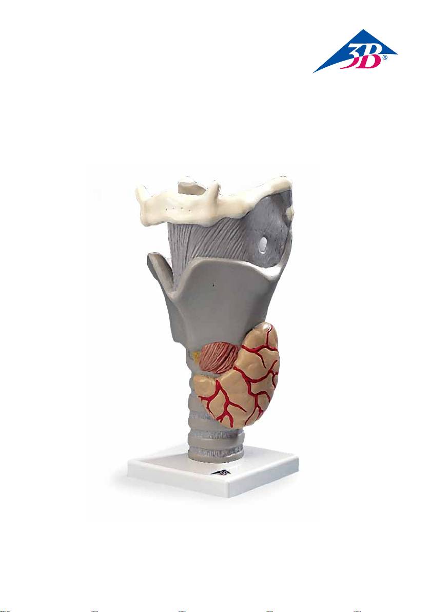
1 Os hyoideum
2 Cartilago thyroidea
3 Cartilago cricoidea
4 Epiglottis
5 Cartilago arytenoidea
6 Glandula thyroidea
7 Glandulae parathyroideae
3

®
Functional Larynx,
English
2.5 times full-size
This model shows the structure of the larynx and the relative position to the larynx of the thyroid gland, of
which the left lobe is presented. The larynx is one of the respiratory organs. The epiglottis can be moved
to demonstrate how the epiglottis closes down over the aperture of the larynx during swallowing, sealing
off the lower respiratory tract. The larynx is furthermore used to generate the voice. The flexible arytenoid
cartilage and vocal chords enable impressive demonstrations of how the true glottis dilates and constricts
during movements of the cricoarytenoid joint.
1 Hyoid bone
2 Thyroid cartilage
3 Cricoid cartilage
4 Epiglottis
5 Arytenoid carilage
6 Thyroid gland
7 Parathyroid glands
4

®
Deutsch
Funktions-Kehlkopf,
2,5-fache Größe
Dieses Modell zeigt den Aufbau des Kehlkopfes sowie die Lage der Schilddrüse, deren linker Lappen darge-
stellt ist, zum Kehlkopf. Der Kehlkopf gehört zu den Atmungsorganen. Durch den beweglichen Kehldeckel
lässt sich anschaulich demonstrieren, wie sich der Kehldeckel beim Schluckakt über den Kehlkopfeingang
legt und somit den Zugang zu den unteren Luftwegen verschließt. Zudem dient der Kehlkopf der
Tonbildung. Durch die Beweglichkeit der Stellknorpel und der Stimmbänder kann eindrucksvoll gezeigt
werden, wie sich die Stimmritze bei Betätigung des Drehgelenkes (Articulatio cricoarytenoidea) erweitert
bzw. verengt.
1 Zungenbein
2 Schildknorpel
3 Ringknorpel
4 Kehldeckel
5 Stellknorpel
6 Schilddrüse
7 Nebenschilddrüsen
5

®
Laringe funcional,
Español
2,5 veces su tamaño natural
Este modelo muestra la estructura de la laringe al igual que la posición de la glándula tiroides, incluyendo
la representación de su lóbulo izquierdo, con respecto a la laringe. La laringe forma parte del aparato
respiratorio. Por medio de la epiglotis móvil se puede demostrar ilustrativamente la manera en que ésta se
coloca por encima de la entrada de la laringe, durante el acto de la deglución, bloqueando de esta manera
el acceso a las vías respiratorias inferiores. Por otra parte, la laringe sirve para la emisión de sonidos.
Gracias a la movilidad del cartílago aritenoides y de las cuerdas vocales se puede mostrar de manera im-
presionante cómo la glotis se expande o se contrae por el accionamiento de la articulación cricoaritenoidea.
1 Hueso hioides
2 Cartílago tiroides
3 Cartílago cricoides
4 Epiglotis
5 Cartílago aritenoides
6 Glándula tiroides
7 Glándula paratiroidea
6

®
Français
Larynx fonctionnel,
agrandi 2,5 fois
Ce modèle illustre la structure du larynx ainsi que la position de la glande thyroïde (dont le lobe gauche est
représenté) par rapport au larynx. Le larynx fait partie de l’appareil respiratoire. La mobilité de l’épiglotte
mobile permet de démontrer parfaitement comment cette dernière se rabat sur l’entrée du larynx, la glot-
te, lors de la déglutition en fermant ainsi les voies respiratoires inférieures. Le larynx sert en outre à la for-
mation de la voix. Grâce à la mobilité du cartilage aryténoïde et des cordes vocales, offerte par ce modèle,
il est possible de démontrer de manière impressionnante comment la fente glottique s’élargit ou se rétrécit
en actionnant l’articulation crico-aryténoïdienne.
1 Os hyoïde
2 Cartilage thyroïde
3 Cartilage cricoïde
4 Épiglotte
5 Cartilage aryténoïde
6 Glande thyroïde
7 Glandes parathyroïdes
7

®
Laringe funcional,
Português
2.5 vezes o tamanho natural
Este modelo mostra a estrutura da laringe assim como a posição da tiróide cujo lobo esquerdo está repre-
sentado perto da laringe. A laringe pertence ao grupo dos órgãos respiratórios. Através da tampa móvel da
laringe pode-se demonstrar de forma visível como no ato de deglutir a tampa da laringe coloca-se por cima
da entrada da laringe e assim fecha a passagem para as vias respiratórias inferiores. Além disso a laringe
serve para a formação de sons. Por meio da mobilidade dos cartilagens de ajuste e das cordas vocais pode
ser mostrado de forma impressionante como as fendas vocais diminuem e aumentam em função da ação
da articulação cricoaritenóidea.
1 Osso hióide
2 Cartilagem tireóidea
3 Cartilagem cricóide
4 Epiglote
5 Cartilagem aritenóide
6 Glândula tireóidea
7 Glândula paratireóidea
8

®
Italiano
Laringe, modello funzionale,
ingrandito 2,5 volte
Questo modello mostra la struttura della laringe e la posizione della tiroide, di cui è rappresentato il lobo
sinistro, rispetto alla laringe. La laringe fa parte degli organi respiratori. Con l’epiglottide mobile si può
dimostrare chiaramente come l’epiglottide durante la deglutizione si chiuda sull’ingresso della laringe,
bloccando l’accesso alle vie respiratorie inferiori. Inoltre la laringe contribuisce alla formazione dei suoni.
Grazie alla mobilità delle aritenoidi e delle corde vocali si può mostrare perfettamente l’allargamento e il
restringimento della glottide con l’attivazione dell’articolazione cricoaritenoidea (Articulatio cricoaryteno-
idea).
1 Osso ioide
2 Cartilagine tiroidea
3 Cartilagine cricoidea
4 Epiglottide
5 Cartilagine aritenoidea
6 Ghiandola tiroidea
7 Ghiandole paratiroidee
9

喉頭,2.5 倍大・デモ用モデル機能可動型
日本語
このモデルでは喉頭の構造,及び甲状腺の左葉との位置関係を見ることができます。喉頭蓋は可動式なので,
嚥下時に喉頭蓋がどのようにして喉頭口をふさぐのかをデモンストレーションすることができます。また,喉
頭は呼吸器としてだけではなく発声という重要な役割も担っています。このモデルの披裂軟骨と声帯は柔軟性
があるので,輪状披裂関節を動かし,声門が拡張・収縮する様子をデモンストレーションすることが可能です。
1 舌骨
2 甲状軟骨
3 輪状軟骨
4 喉頭蓋
5 披裂軟骨
6 甲状腺
7 上皮小体(副甲状腺)
10

Русский
Функциональная модель гортани,
увеличение в 2,5 раза
На данной модели показано строение гортани и расположение щитовидной железы (представлена левая
доля) относительно гортани. Гортань – это один из органов дыхания. Надгортанник в модели является
подвижной частью для демонстрации того, как он опускается сверху на апертуру гортани во время
глотания, герметично закрывая нижние дыхательные пути. Кроме того, гортань используется для генерации
голоса. Гибкий черпаловидный хрящ и голосовые связки дают возможность получить представление о том,
как сужается и расширяется голосовая щель во время движений в перстнечерпаловидном суставе.
1 Подъязычная кость
2. Щитовидный хрящ
3 Перстневидный хрящ
4 Надгортанник
5 Черпаловидный хрящ
6 Щитовидная железа
7 Паращитовидные железы
中文
功能性喉部
实际大小的2.5倍
这个模型显示了喉的结构,以及甲状腺与喉的相对位置,模型呈现了甲状腺的左叶。喉部是一个呼
吸器官,会厌能够移动,用来证明在进行吞咽的时候,会厌是如何来关闭喉口,从而来封闭下呼吸
道。此外,喉部也用来产生声音,灵活的杓状软骨和声带使得实物教授能够给人深刻的印象,说明
了在环杓关节运动期间,声门裂如何开大和缩小。
1 舌骨
2 甲状软骨
3 环状软骨
4 会厌
5 杓状软骨
6 甲状腺
7 甲状旁腺
11

3B Scientific GmbH
Rudorffweg 8 • 21031 Hamburg • Germany
Tel.: + 49-40-73966-0 • Fax: + 49-40-73966-100
www.3bscientific.com • 3b@3bscientific.com
© Copyright 2006 / 2013 for instruction manual and design of product:
3B Scientific GmbH, Germany
© Copyright 2011 for instruction manual and design of product:
3B Scientific GmbH, Germany
3B Scientific
A w o r l d w i d e g r o u p o f c o m p a n i e s
5001995 G20 (1000271)-02/13-2
