3B Scientific Shoulder Joint with Rotator Cuff - 5 part: инструкция
Раздел: Товары для здоровья
Тип:
Инструкция к 3B Scientific Shoulder Joint with Rotator Cuff - 5 part
A880
(1000176)

2

®
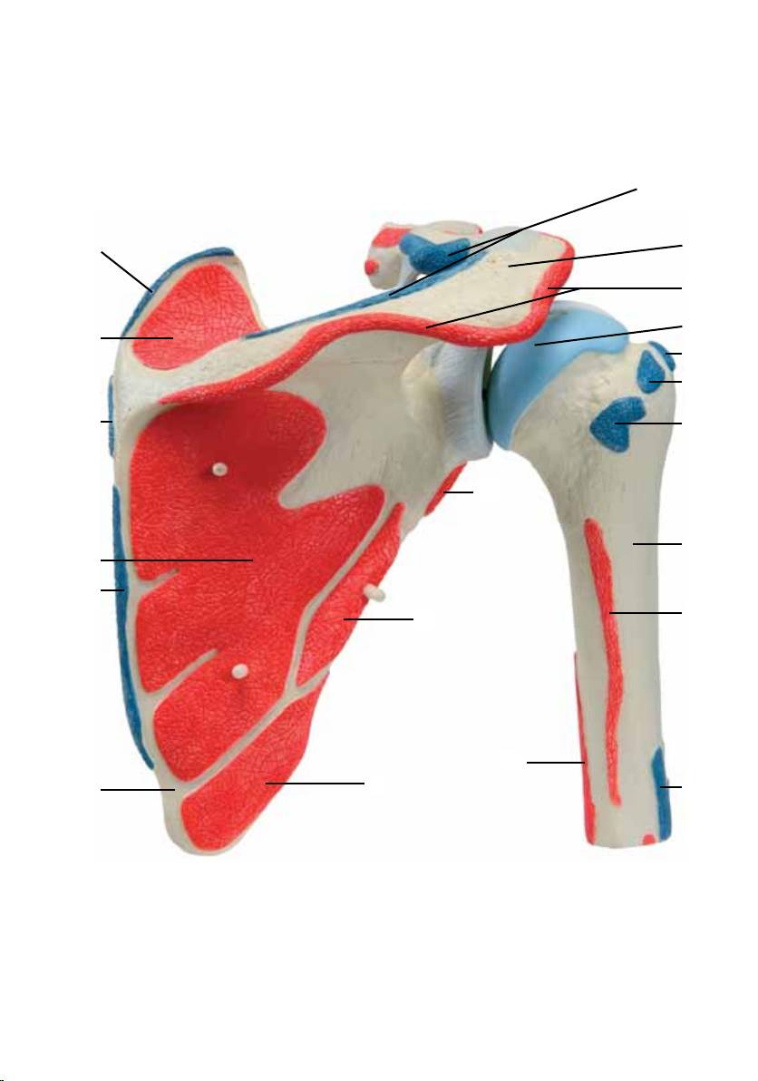
Latin
1 Clavicula
2 M. supraspinatus
3 M. serratus anterior, insertio
4 M. subscapularis
5 M. deltoideus, insertio
6 Humerus
7 M. teres major, insertio
8 M. latissimus dorsi, insertio
9 M. pectoralis major, insertio
10 Caput longum m. bicepitis brachii, tendo
11 M. pectoralis minor, insertio
12 M. coracobrachialis, origo
13 Caput breve m. bicepitis brachii, origo
14 Processus coracoideus
15 Lig. coracoacromiale
16 M. trapezius, insertio
17 Acromion
18 M. deltoideus, origo
19 M. infraspinatus
20 M. teres minor
21 Caput laterale m. tricepitis brachii,origo
22 Caput mediale m. tricepitis brachii, origo
23 M. teres major, origo
24 Scapula
25 M. rhomboideus major, insertio
26 M. rhomboideus minor, insertio
27 Spina scapulae
28 M. levator scapulae, insertio
29 M. subscapularis, insertio
30 Caput humeri
31 Lig. coracoclaviculare, Lig. conoideum
32 M. subscapularis, origo
33 M. supraspinatus, insertio
34 M. infraspinatus, insertio
35 M. teres minor, insertio
36 Caput longum m. tricepitis brachii, origo
37 M. teres minor, origo
38 M. infraspinatus, origo
39 M. supraspinatus, origo
40 M. sternocleidomastoideus, origo
41 M. pectoralis major, origo
42 Lig. coracoclaviculare, Lig. trapezoideum
43 Lig. acromioclaviculare
3

®
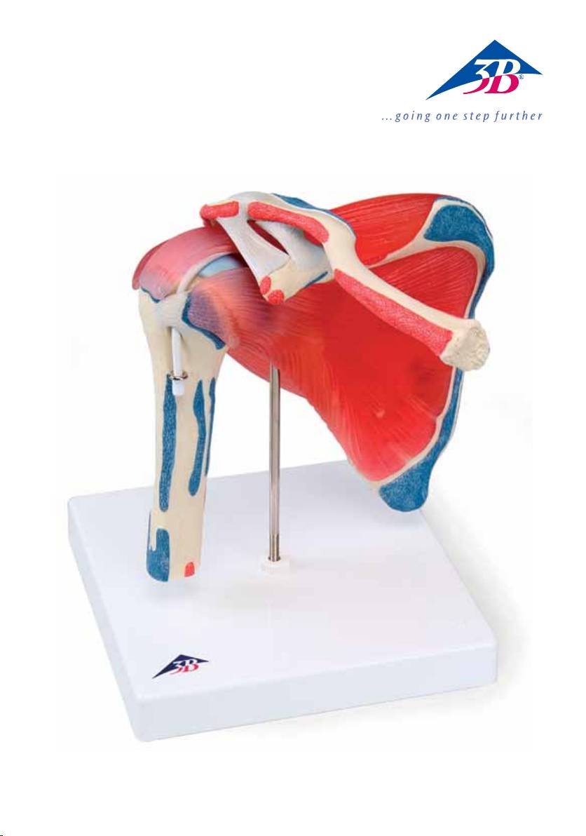
Shoulder Joint with Rotator Cuff
English
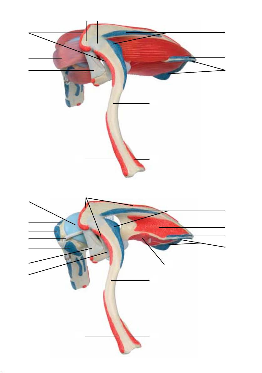
The model shows the shoulder joint and, in addition to the individual muscles of the rotator cuff, the bone
origins and insertions of all shoulder muscles. For didactic reasons, the areas of origin and insertion of the
individual muscles are raised and colored (origin = red; insertion = blue).
The rotator cuff consists of four muscles: subscapularis muscle, supraspinatus muscle, infraspinatus mus-
cle and teres minor muscle. Each muscle is attached to its matching areas of origin and insertion and is
detachable. The muscles are made of a half-transparent, muscle-colored material, so that the correspondi-
ng areas of origin and insertion can be seen even while the muscles are attached.
The muscles of the rotator cuff have the following functions:
• Subscapularis muscle: medial rotation
• Supraspinatus muscle: abduction and lateral rotation
• Infraspinatus muscle: lateral rotation
• Teres minor muscle: lateral rotation and adduction
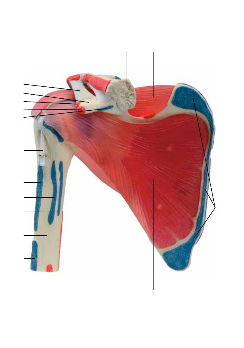
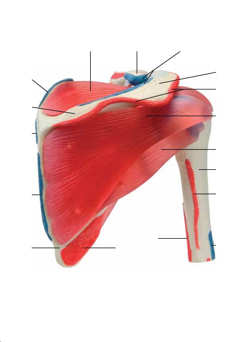
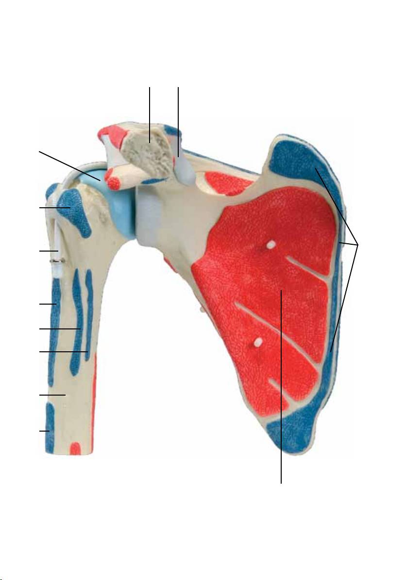
1 Clavicle
38 Infraspinatus muscle, origin
2 Supraspinatus muscle
39 Supraspinatus muscle, origin
3 Serratus anterior muscle, insertion
40 Sternocleidomastoid muscle, origin
4 Subscapularis muscle
41 Pectoralis major muscle, origin
5 Deltoideus muscle, insertion
42 External coracoclavicular ligament
6 Humerus
43 Acromioclavicular ligament
7 Teres major muscle, insertion
8 Latissimus dorsi muscle, insertion
9 Pectoralis major muscle, insertion
10 Long head of biceps brachii muscle, tendon
11 Pectoralis minor muscle, insertion
12 Coracobrachialis muscle, origin
13 Short head of biceps brachii muscle, origin
14 Coracoid process
15 Coracoacromial ligament
16 Trapezius muscle, insertion
17 Acromion
18 Deltoideus muscle, origin
19 Infraspinatus muscle
20 Teres minor muscle
21 Lateral head of triceps brachii muscle, origin
22 Medial head of triceps brachii muscle, origin
23 Teres major muscle, origin
24 Scapula
25 Rhomboideus major muscle, insertion
26 Rhomboideus minor muscle, insertion
27 Spine of scapula
28 Levator scapulae muscle, insertion
29 Subscapularis muscle, insertion
30 Head of humerus
31 Internal coracoclavicular ligament
32 Subscapularis muscle, origin
33 Supraspinatus muscle, insertion
34 Infraspinatus muscle, insertion
35 Teres minor muscle, insertion
36 Long head of triceps brachii muscle, origin
37 Teres minor muscle, origin
4

®
Deutsch Schultergelenk mit Rotatorenmanschette
Das Modell zeigt das Schultergelenk und neben den einzelnen Muskeln der Rotatorenmanschette die
Knochenursprünge und -ansätze der gesamten Schultermuskulatur. Aus didaktischen Gründen sind die
Ursprungs- und Ansatzflächen der einzelnen Muskeln erhöht und farbig (Ursprung = rot ; Ansatz = blau)
dargestellt.
Die Rotatorenmanschette besteht aus vier Muskeln: M. subscapularis (Unterschulterblattmuskel), M.
supraspinatus (Obergrätenmuskel), M. infraspinatus (Untergrätenmuskel) und M. teres minor (Kleiner
Rundmuskel). Die einzelnen Muskeln werden auf ihren jeweiligen Ursprungs- und Ansatzflächen aufge-
steckt und sind somit abnehmbar. Sie sind aus halbtransparentem, muskelfarbigen Material, wodurch die
jeweiligen Ursprungs- und Ansatzflächen auch bei aufgesteckter Muskulatur zu erkennen sind.
Die Muskeln der Rotatorenmanschette haben folgende Funktionen:
• M. subscapularis: Innenrotation
• M. supraspinatus: Abduktion und Außenrotation
• M. infraspinatus: Außenrotation
• M. teres minor: Außenrotation und Adduktion
1 Schlüsselbein
30 Oberarmkopf
2 Obergrätenmuskel
31 Hinterer Teil des Bindegewebsbandes zwischen
3 Vorderer Sägemuskel, Ansatz
Rabenschnabelfortsatz und Schlüsselbein
4 Unterschulterblattmuskel
32 Unterschulterblattmuskel,Ursprung
5 Deltamuskel, Ansatz
33 Obergrätenmuskel, Ansatz
6 Oberarmknochen
34 Untergrätenmuskel, Ansatz
7 Großer Rundmuskel, Ansatz
35 Kleiner Rundmuskel, Ansatz
8 Breiter Rückenmuskel, Ansatz
36 Langer Kopf des dreiköpfigen Oberarmmuskels,
9 Großer Brustmuskel, Ansatz
Ursprung
10 Langer Kopf des zweiköpfigen
37 Kleiner Rundmuskel, Ursprung
Oberarmmuskels, Sehne
38 Untergrätenmuskel, Ursprung
11 Kleiner Brustmuskel, Ansatz
39 Obergrätenmuskel, Ursprung
12 Hakenarmmuskel, Ursprung
40 Kopfwendemuskel, Ursprung
13 Kurzer Kopf des zweiköpfigen
41 Großer Brustmuskel, Ursprung
Oberarmmuskels, Ursprung
42 Vorderer Teil des Bindegewebsbandes zwischen
14 Rabenschnabelfortsatz
Rabenschnabelfortsatz und Schlüsselbein
15 Bindegewebsband zwischen
43 Bindegewebsband zwischen Schulterhöhe und
Rabenschnabelfortsatz und Schulterhöhe
Schlüsselbein
16 Kappenmuskel, Ansatz
17 Schulterhöhe
18 Deltamuskel, Urspung
19 Untergrätenmuskel
20 Kleiner Rundmuskel
21 Seitlicher Kopf des dreiköpfigen
Oberarmmuskels, Ursprung
22 Mittlerer Kopf des dreiköpfiger
Oberarmmuskel, Ursprung
23 Großer Rundmuskel, Ursprung
24 Schulterblatt
25 Großer Rautenmuskel, Ansatz
26 Kleiner Rautenmuskel, Ansatz
27 Schulterblattgräte
28 Schulterblattheber, Ansatz
29 Unterschulterblattmuskel, Ansatz
5
1 2
15
14
13
12
11
10
9
8
3
7
6
5
4
6
2 1 16
17
28
18
27
19
26
20
6
21
25
22
5
24 23
7
1 31
30
29
3
10
9
8
7
6
5
32
8
16
17
28
18
30
39
33
34
26
35
36
6
38
25
21
37
22
23
5
24
9
17 43
18
16
28
2
15
3
1
41
40
18
30
16
34
39
33
28
10
29
3
15
32
42
1
41
40
10

®
Español
Articulación del hombro
y manguito rotatorio
El modelo muestra la articulación del hombro y cada uno de los músculos del manguito rotatorio, junto a
sus orígenes e inserciones musculares. Por razones didácticas, los orígenes y las inserciones de cada mús-
culo están realzadas y coloreadas (origen= rojo; inserción= azul). El manguito rotatorio está formado por
cuatro músculos: músculo subescapular, músculo supraespinoso, músculo infraespinoso y músculo redon-
do menor. Cada músculo está fijado a sus correspondientes orígenes e inserciones y se puede desmontar.
El material utilizado es semitransparente coloreado, lo que permite reconocer igualmente los orígenes e
inserciones de los músculos una vez desmontados. Los músculos del manguito rotatorio tienen las fun-
ciones siguientes:
• M. subescapular: rotación interna
• M. supraespinoso: abducción y rotación externa
• M. infraespinoso: rotación externa
• M. redondo menor: rotación externa y aducción.
1 Clavícula
33 M. supraespinoso, inserción
2 M. supraespinoso
34 M. infraespinoso, inserción
3 M. serrato mayor, inserción
35 M. redondo menor, inserción
4 M. subescapular
36 Cabeza larga del músculo tríceps braquial,
5 M. deltoides, inserción
origen
6 Húmero
37 M. redondo menor, origen
7 M. redondo mayor, inserción
38 M. infraespinoso, origen
8 M. dorsal ancho, inserción
39 M. supraespinoso, origen
9 M. pectoral mayor, inserción
40 M. estrenocleidomastoideo, orígen
10 Cabeza larga del músculo bíceps braquial,
41 M. pectoral mayor, origen
tendón
42 Lig. coracoclavicular, lig. trapezoideo
11 M. pectoral menor, inserción
43 Lig. acromioclavicular
12 M. coracobraquial, origen
13 Cabeza corta del músculo bíceps braquial,
origen
14 Apófisis coracoides
15 Lig. coracoacromial
16 M. trapecio, inserción
17 Acromion
18 M. deltoides, origen
19 M. infraespinoso
20 M. redondo menor
21 Cabeza lateral del músculo tríceps braquial,
origen
22 Cabeza mediana del músculo tríceps braquial,
origen
23 M. redondo mayor, origen
24 Escápula
25 M. romboideo mayor, inserción
26 M. romboideo menor, inserción
27 Espina escapular
28 M. elevador de la escápula, inserción
29 M. subescapular, inserción
30 Cabeza del húmero
31 Lig. coracoclavicular, lig. Conoide
32 M. subescapular, origen
11

®
Articulation de l‘épaule avec
FrançaisName
manchette des rotateurs
Le modèle montre l‘articulation de l‘épaule et en plus des différents muscles de la manchette des rota-
teurs, les origines et les insertions des os de la musculature complète de l‘épaule. Pour des raisons didac-
tiques, les surfaces d‘origine et d‘insertion des différents muscles sont représentées de façon surélevée et
colorée (origine = rouge ; insertion = bleu). La manchette des rotateurs est composée de quatre muscles :
M. subscapularis (muscle sous-scapulaire), M. supraspinatus (muscle sus-épineux), M. infraspinatus (muscle
sous-épineux) et M. teres minor (muscle petit rond). Les différents muscles sont emboîtés sur leur surfaces
d‘origine et d‘insertion correspondantes et sont, par conséquent, amovibles. Ils sont fabriqués en matériau
semi-transparent ayant la teinte des muscles, permettant de reconnaître les différentes surfaces d‘origine
et d‘insertion, même sur la musculature emboîtée. Les muscles de la manchette des rotateurs ont les fonc-
tions suivantes :
• M. subscapularis : rotation interne
• M. supraspinatus : abduction et rotation externe
• M. infraspinatus : rotation externe
• M. teres minor : rotation externe et adduction
1 Clavicule
31 Ligament coraco-claviculaire, ligament conoïde
2 Muscle supra-épineux
32 Muscle subscapulaire, origine
3 Muscle dentelé antérieur, insertion
33 Muscle supra-épineux, insertion
4 Muscle subscapulaire
34 Muscle infra-épineux, insertion
5 Muscle deltoïde, insertion
35 Muscle petit rond, insertion
6 Humérus
36 Longue tête du muscle de l‘humérus à trois
7 Muscle grand rond, insertion
têtes, origine
8 Muscle grand dorsal, insertion
37 Muscle petit rond, origine
9 Muscle grand pectoral, insertion
38 Muscle infra-épineux, origine
10 Longue tête du muscle de l‘humérus à deux
39 Muscle supra-épineux, origine
têtes, tendon
40 M. sterno-cléido-mastoïdien, origine
11 Muscle petit pectoral, insertion
41 Muscle grand pectoral, origine
12 Muscle coraco-brachial, origine
42 Ligament coraco-claviculaire, ligament
13 Tête courte du muscle de l‘humérus à deux
trapézoïde
têtes, origine
43 Ligament acromio-claviculaire
14 Processus coracoïde
15 Ligament coraco-acromial
16 Muscle trapèze, insertion
17 Acromion
18 Muscle deltoïde, origine
19 Muscle infra-épineux
20 Muscle petit rond
21 Tête latérale du muscle de l‘humérus à trois
têtes, origine
22 Tête centrale du muscle de l‘humérus à trois
têtes, origine
23 Muscle grand rond, origine
24 Scapula
25 Muscle grand rhomboïde, insertion
26 Muscle petit rhomboïde, insertion
27 Epine de l‘omoplate
28 Muscle élévateur de la scapula, insertion
29 Muscle subscapulaire, insertion
30 Tête de l‘humérus
12

®
Português Name
Articulação do ombro
com manga de rotores
Manga de rotatores, origens e inserções de toda a musculatura do ombro. Por razões didáticas, as superfí-
cies das origens e das inserções de cada músculo estão elevadas e representadas em cores (origens = ver-
melho; inserções = azul). A manga de rotores consiste em quatro músculos: subescapular, supraespinhal,
infraespinhal e teres menor. Cada músculo é encaixado nas suas origens e inserções correspondentes e são
portanto removíveis. Eles são feitos de um material semi-transparente da cor dos músculos, sendo que
mesmo com os músculos encaixado ainda pode-se reconhecer as origens e as inserções.
Os músculos da manga de rotores têm as seguintes funções:
• M. subescapular: rotação interna
• M. supraespinhal: abdução e rotação externa
• M. infraespinhal: rotação externa
• M. teres menor: rotação externa e abdução
1 Clavícula
34 Músculo infra-espinhoso, inserção
2 Músculo supra-espinhoso
35 Músculo redondo menor, inserção
3 Músculo serrátil anterior, inserção
36 Cabeça longa do músculo tríceps do braço,
4 Músculo subescapular
origem
5 Músculo deltóide, inserção
37 Músculo redondo menor, origen
6 Úmero
38 Músculo infra-espinhoso, origen
7 Músculo redondo maior, inserção
39 Músculo supra-espinhoso, origen
8 Músculo grande dorsal, inserção
40 Músculo sternocleidomastóide, origem
9 Músculo grande peitoral, inserção
41 Músculo grande peitoral, origen
10 Cabeça longa do músculo bíceps do braço,
42 Ligamento coracoclavicular, ligamento
tendão
trapezóide
11 Músculo peitoral menor, inserção
43 Ligamento acromioclavicular
12 Músculo coracobraquial, origen
13 Cabeça curta do músculo bíceps do braço,
origem
14 Processo coracóide
15 Ligamento coracoacromial
16 Músculo trapézio, inserção
17 Acrômio
18 Músculo deltóide, origen
19 Músculo infra-espinhoso
20 Músculo redondo menor
21 Cabeça lateral do músculo tríceps do braço,
origem
22 Cabeça mediana do músculo tríceps do braço,
origem
23 Músculo redondo maior, origen
24 Escápula
25 Músculo rombóide maior, inserção
26 Músculo rombóide menor, inserção
27 Espinha da escápula
28 Músculo elevador da escápula, inserção
29 Músculo subescapular, inserção
30 Cabeça do braço
31 Ligamento coracoclavicular, ligamento conóide
32 Músculo subescapular, origen
33 Músculo supra-espinhoso, inserção
13

®
Articolazione della spalla
Italiano
con Cuffia dei rotatori
Questo modello mostra l’articolazione della spalla con i muscoli della cuffia dei rotatori e con le origini e
attaccature delle ossa di tutta la muscolatura della spalla. Per ragioni didattiche le aree di origine e attac-
catura dei muscoli sono rappresentate in rilievo e di colore diverso (origini in rosso e attaccature in blu).
La cuffia dei rotatori è composta da quattro muscoli: m. sottoscapolare, m. sopraspinato, m. infraspinato e
m. rotondo piccolo. I muscoli sono inseriti nelle relative aree di origine e attaccatura, e sono quindi stacca-
bili. Sono in materiale semitrasparente, color muscolo, per rendere riconoscibili origini e attaccature anche
quando la muscolatura è in posizione.
I muscoli della cuffia dei rotatori hanno le seguenti funzioni:
• m. sottoscapolare: rotazione interna
• m. sopraspinato: abduzione e rotazione esterna
• m. infraspinato: rotazione esterna
• m. rotondo piccolo: rotazione esterna e adduzione
1 Clavicola
33 M. sopraspinato, attaccatura
2 M. sopraspinato
34 M. infraspinato, attaccatura
3 M. grande dentato, attaccatura
35 M. rotondo piccolo, attaccatura
4 M. sottoscapolare
36 Capo lungo del muscolo brachiale a tre capi,
5 M. deltoide, attaccatura
origine
6 Omero
37 M. rotondo piccolo, origine
7 M. rotondo grande, attaccatura
38 M. infraspinato, origine
8 M. larghissimo del dorso, attaccatura
39 M. sopraspinato, origine
9 M. pettorale grande, attaccatura
40 M. sternocleidomastoideo, origine
10 Capo lungo del muscolo brachiale a due capi,
41 M. pettorale grande, origine
tendine
42 Leg. coracoclavicolare, leg trapezoideo
11 M. pettorale piccolo, attaccatura
43 Leg. acromioclavicolare
12 M. coracobrachiale, origine
13 Capo corto del muscolo brachiale a due capi,
origine
14 Processo coracoideo
15 Leg. coraco-acromiale
16 M. trapezio, attaccatura
17 Acromion
18 M. deltoide, origine
19 M. infraspinato
20 M. rotondo piccolo
21 Capo laterale del muscolo brachiale a tre capi,
origine
22 Capo medio del muscolo brachiale a tre capi,
origine
23 M. rotondo grande, origine
24 Scapola
25 M. romboide grande, attaccatura
26 M. romboide piccolo, attaccatura
27 Spina dell’omoplata
28 M. elevatore della scapola, attaccatura
29 M. sottoscapolare, attaccatura
30 Testa dell’omero
31 Leg. coracoclavicolare, leg. conoideo
32 M. sottoscapolare, origine
14

日本語
ローテーターカフ付肩関節モデル
このモデルでは,肩関節やローテーターカフ(回旋筋腱板)の4つの筋に加え,肩部の筋全ての起始/停止を
見ることができます。起始は赤
,停止は青に色付けして表現しています。
ローテーターカフは
,肩甲下筋,棘上筋,棘下筋,小円筋の4つの筋からなります。このモデルでは,各筋の
取り外しが可能で
,それぞれの起始/停止も明示しています。筋肉は半透明なので,付けたままの状態でも符
合する起始/停止を確認することができます。
このモデルでは
,次の動きを表現可能です。
• 肩甲下筋:内側回旋
• 棘 上 筋:外転と外側回旋
• 棘 下 筋:外側回旋
• 小 円 筋:外側回旋と内転
1 鎖骨
36 上腕三頭筋の長頭,起始
2 棘上筋
37 小円筋,起始
3 前鋸筋,停止
38 棘下筋,起始
4 肩甲下筋
39 棘上筋,起始
5 三角筋,停止
40 胸鎖乳突筋,起始
6 上腕骨
41 大胸筋,起始
7 大円筋,停止
42 菱形靭帯
8 広背筋,停止
43 肩鎖靭帯
9 大胸筋,停止
10 上腕二頭筋の長頭,腱
11 小胸筋,停止
12 鳥口腕筋,起始
13 上腕二頭筋の短頭,起始
14 鳥口突起
15 鳥口肩峰靭帯
16 僧帽筋,停止
17 肩峰
18 三角筋,起始
19 棘下筋
20 小円筋
21 上腕三頭筋の外側頭,起始
22 上腕三頭筋の内側頭,起始
23 大円筋,起始
24 肩甲骨
25 大菱形筋,停止
26 小菱形筋,停止
27 肩甲棘
28 肩甲挙筋,停止
29 肩甲下筋,停止
30 上腕骨頭
31 円錐靭帯
32 肩甲下筋,起始
33 棘上筋,停止
34 棘下筋,停止
35 小円筋,停止
15
A880__.indd 15 05.5.7 10:03:29 AM

®
РусскийВращающая манжета плеча
Модель показывает плечевой сустав, а также, помимо отдельных мышц вращающей манжеты плеча,
точки начала и прикрепления к костям всех мышц плеча. Для большей наглядности обучения области
начала и прикрепления отдельных мышц сделаны выпуклыми и обозначены цветом (начало – красным,
прикрепление – синим). Вращающая манжета плеча состоит из четырех мышц: подлопаточной
мышцы, надостной мышцы, подостной мышцы и малой круглой мышцы. Каждая мышца прикреплена
к соответствующим областям начала и прикрепления, их можно отсоединять. Мышцы изготовлены из
полупрозрачного, окрашенного в цвет мышц материала, поэтому соответствующие области начала и
прикрепления можно увидеть даже, когда мышцы присоединены.
Мышцы вращающей манжеты плеча выполняют следующие функции:
• Подлопаточная мышца: медиальная ротация
• Надостная мышца: приведение и латеральная ротация
• Подостная мышца: латеральная ротация
• Малая круглая мышца: латеральная ротация и отведение
1 Ключица
30 Головка плечевой кости
2 Надостная мышца
31 Внутренняя клювовидно-ключичная связка
3 Передняя зубчатая мышца, место
32 Подлопаточная мышца, начало
прикрепления
33 Надостная мышца, место прикрепления
4 Подлопаточная мышца
34 Подостная мышца, место прикрепления
5 Дельтовидная мышца, место прикрепления
35 Малая круглая мышца, место прикрепления
6 Плечевая кость
36 Длинная головка трехглавой мышцы плеча,
7 Большая круглая мышца, место прикрепления
начало
8 Широчайшая мышца спины, место
37 Малая круглая мышца, начало
прикрепления
38 Подостная мышца, начало
9 Большая грудная мышца, место прикрепления
39 Надостная мышца, начало
10 Длинная головка двуглавой мышцы плеча,
40 Грудино-ключично-сосцевидная мышца,
сухожилие
начало
11 Малая грудная мышца, место прикрепления
41 Большая грудная мышца, начало
12 Клювовидно-плечевая мышца, начало
42 Наружная клювовидно-ключичная связка
13 Короткая головка двуглавой мышцы плеча,
43 Акромиально-ключичная связка
начало
14 Клювовидный отросток
15 Клювовидно-акромиальная связка
16 Трапециевидная мышца, место прикрепления
17 Акромион
18 Дельтовидная мышца, начало
19 Подостная мышца
20 Малая круглая мышца
21 Латеральная головка трехглавой мышцы
плеча, начало
22 Медиальная головка трехглавой мышцы плеча,
начало
23 Большая круглая мышца, начало
24 Лопатка
25 Большая ромбовидная мышца, место
прикрепления
26 Малая ромбовидная мышца, место
прикрепления
27 Ость лопатки
28 Мышца, поднимающая лопатку, место
прикрепления
29 Подлопаточная мышца, место прикрепления
16

®
中文 肩袖
该模型显示肩关节,以及组成肩袖的各块肌肉,同时该模型上还清晰的显示了各块肌肉的起止点。
为了便于用户使用,我们特意使用了不同的颜色来标示出肌肉的起止点(起点:红色;止点:蓝
色)。
肩袖由4块肌肉组成:肩胛下肌;冈上肌;冈下肌;以及小圆肌。每块肌肉均有清晰的相应起止
点,并且可以从模型上拆卸下来。肌肉是由半透明材料制成,与人体肌肉颜色相似,因此在该模型
上,即使肌肉是附着的,用户也可以清晰的看见肌肉的起止点。肩袖的肌肉有以下的功能:
·肩胛下肌:内旋;
·冈上肌:外展,外旋;
·冈下肌:外旋;
·小圆肌:外旋内收。
1 锁骨
42 外侧喙锁韧带
2 冈上肌
43 肩锁韧带
3 前锯肌,止点
4 肩胛下肌
5 三角肌,止点
6 肱骨
7 大圆肌,止点
8 背阔肌,止点
9 胸大肌,止点
10 肱二头肌长头,肌腱;
11 胸小肌,止点
12 喙肱肌,起点;
13 肱二头肌短头,起点;
14 喙突
15 喙肩韧带
16 斜方肌,止点
17 肩峰
18 三角肌,起点
19 冈下肌
20 小圆肌
21 肱三头肌长头,起点
22 肱三头肌内侧头,起点
23 大圆肌,起点
24 肩胛骨
25 大菱形肌,止点
26 小菱形肌,止点
27 肩胛骨脊
28 肩胛提肌,止点
29 肩胛下肌,止点
30 肱骨头
31 内侧喙锁韧带
32 肩胛下肌,起点
33 冈上肌,止点
34 冈下肌,止点
35 小圆肌,止点
36 肱三头肌长头,起点
37 小圆肌,起点
38 冈下肌,起点
39 冈上肌,起点
40 胸锁乳突肌,起点
41 胸大肌,起点
17

®
18

®
19

A880_1000176-07/12-2
®
3B S C I E NT I F I C
P R O D U C T S
3B Scientific GmbH
Rudorffweg 8 • 21031 Hamburg • Germany
Tel.: + 49-40-73966-0 • Fax: + 49-40-73966-100
www.3bscientific.com • 3b@3bscientific.com
© Copyright 2005 / 2012 for instruction manual and design of product:
3B Scientific GmbH, Germany
