3B Scientific Gastric Band Model: инструкция
Раздел: Товары для здоровья
Тип:
Инструкция к 3B Scientific Gastric Band Model
… g o i n g o n e s t e p f u r t h e r
1012787

2

®
Latin
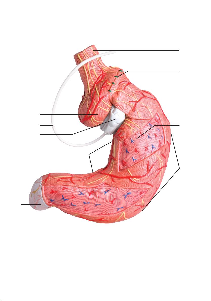
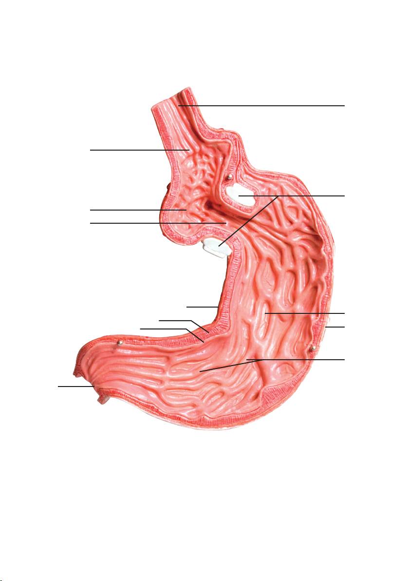
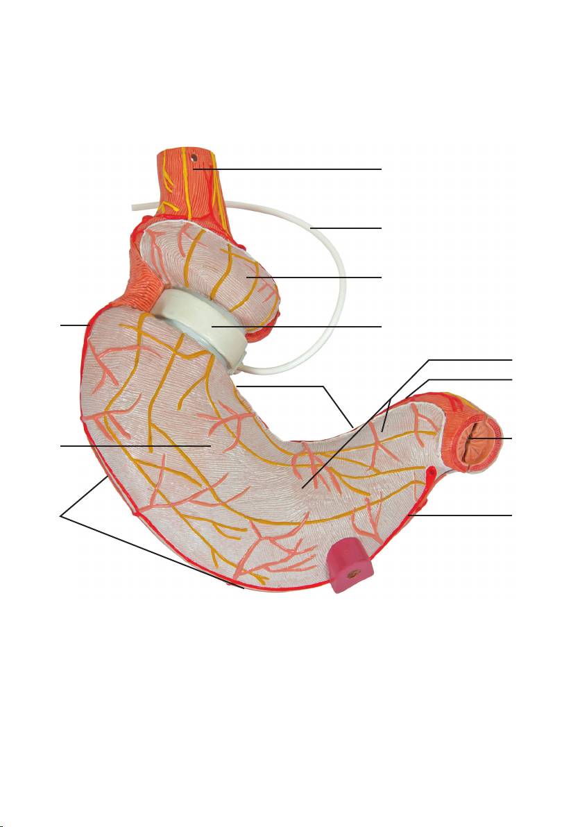
1 Oesophagus
2 Sutura
3 Corpus gastricae
4 Curvatura major
5 Pylorus
6 Curvatura minor
7 Adjustable gastric band
8 Connecting tube
9 Corpus gastricae (Pouch)
10 Inflateable band
11 Plicae gastricae
12 Tunica mucosa
13 Tunica muscularis
14 Stoma
15 Cardia
16 A. gastroomentalis sinistra
17 A. gastroomentalis dextra
18 A. gastrica dextra
19 Peritoneum viscerale
3

®
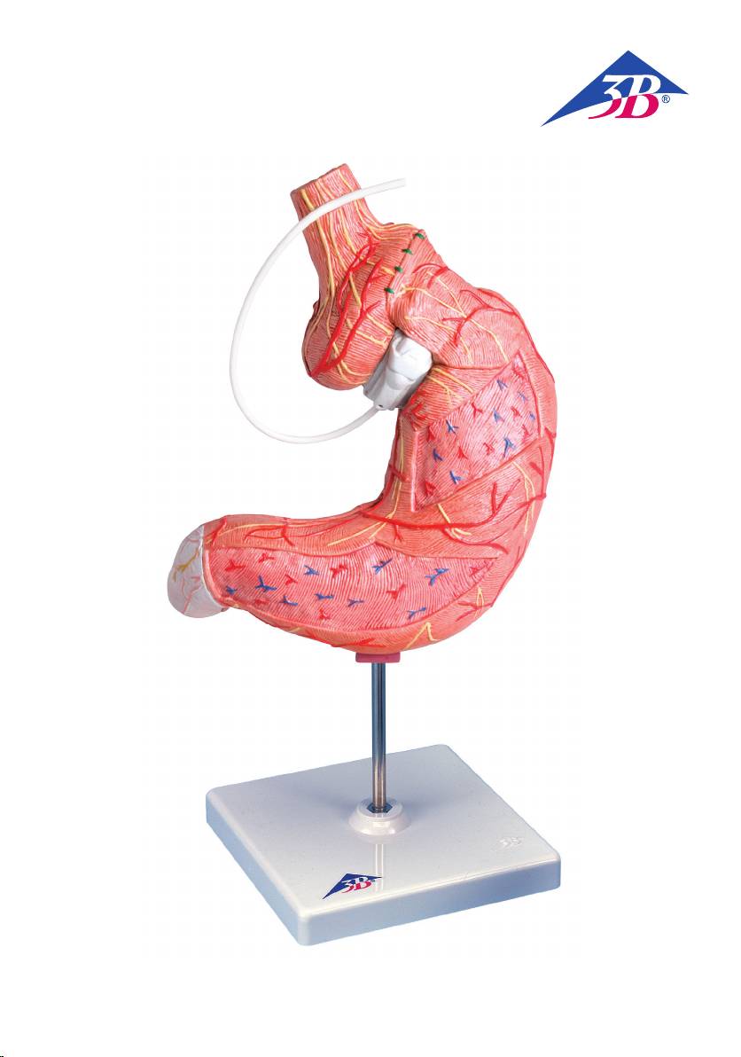
Gastric Band Model. 2 part
English
This model shows the position of an implanted gastric band. The gastric band is placed around the stoma-
ch in such a way that it divides the stomach into a small pouch and a larger stomach section. The resulting
narrow passage (stoma) slows down the passage of food and the stomach wall becomes tight more quickly,
leading to a feeling of fullness. The width of the passageway can be varied by adjusting the amount of
liquid in the inflatable balloon.
1 Oesophagus, Esophagus
2 Anti-slip stitches
3 Larger stomach portion
4 Greater curvature
5 Pylorus
6 Lesser curvature
7 Adjustable gastric band
8 Connecting tube
9 Small stomach pouch
10 Inflateable band
11 Rugal folds
12 Mucosa
13 Muscular layer
14 Stoma
15 Cardia
16 Left gastro-omental artery
17 Right gastro-omental artery
18 Right gastric artery
19 Visceral peritoneum
4

®
Deutsch
Magenband
Dieses Modell zeigt die Lage eines eingesetzten Magenbandes. Das Magenband wird so um den Magen
gelegt, dass es diesen in einen kleinen Vormagen (Pouch) und einen größeren Restmagen unterteilt. Die
dadurch entstehende Enge (Stoma) verzögert die Nahrungspassage und führt zu einer früheren Dehnung
der Magenwand und daraus folglich einsetzendem Sättigungsgefühl. Über den mit Flüssigkeit befüllbaren
Ballon kann die Enge variabel eingestellt werden.
1 Speiseröhre
2 Anti-Slipping Nähte
3 Restmagen
4 Große Kurvatur
5 Magenpförtner
6 Kleine Kurvatur
7 Magenband
8 Schlauchsystem
9 Vormagen (Pouch)
10 Mit Flüssigkeit befüllbarer Ballon
11 Magenfalten
12 Schleimhaut
13 Muskelschicht
14 Enge (Stoma)
15 Mageneingang
16 Linke Schlagader des Magens und des großen Netzes
17 Rechte Schlagader des Magens und des großen Netzes
18 Rechte Magenschlagader
19 Bauchfell
5

®
EspañolModelo de banda gástrica
Este modelo nos muestra la posición de una banda gástrica implantada.
La banda gástrica se coloca alrededor del estómago, de tal modo que lo divide en una bolsa estomacal
anterior y el fondo del estómago restante. El estrechamiento así resultante, (estoma) retrasa el paso de los
alimentos y produce una más rápida expansión de la pared del estómago y la sensación de saciedad resul-
tante. A través del balón gástrico que puede llenarse de líquido, el estrechamiento puede regularse.
1 Esófago
2 Sutura que impide el desplazamiento de la banda
3 Fondo gástrico
4 Curvatura mayor
5 Píloro
6 Curvatura menor
7 Banda gástrica ajustable
8 Sistema de tubos
9 Bolsa pequeña del estómago
10 Balón rellenable con solución salina
11 Pliegues estomacales
12 Mucosa
13 Túnica muscular
14 Estoma
15 Cardias
16 Arteria gastro-epiploica izquierda
17 Arteria gástro-epiploica derecha
18 Arteria gástrica derecha
19 Peritoneo
6

®
Français
Modèle Bande Gastrique
La bande gastrique est constitué d’un anneau gastrique ajustable que l’on place autour de l’estomac, de
manière à le diviser en un pré-estomac (poche) et un plus grand estomac résiduel. L’étroitesse ainsi obte-
nue (stomie) ralentit le passage des aliments et entraîne ainsi une extension plus prématurée de la paroi
gastrique suivie d’une sensation de satiété. L’étroitesse peut être réglée à l’aide du ballon pouvant être
rempli de liquide.
1 Œsophage
2 Sutures anti-glissement
3 Estomac résiduel
4 Grande courbure
5 Pylore
6 Petite courbure
7 Bande gastrique
8 Système tubulaire
9 Pré-estomac (poche)
10 Ballon pouvant être rempli de liquide
11 Replis gastriques
12 Muqueuse
13 Couche musculaire
14 Stomie
15 Cardia
16 Artère gastro-oesophagienne gauche
17 Artère gastro-oesophagienne droite
18 Artère gastrique droite
19 Péritoine
7
1
2
9
8
3
7
6
4
5
8
1
15
10
9
14
6
3
13
4
12
11
5
9
1
8
9
16
7
19
6
18
5
3
4
17
10

®
Português
Modelo de banda gástrica, 2 partes
Este modelo mostra a posição de uma banda gástrica implantada. A banda gástrica é colocada à volta do
estômago, de uma forma que o divide numa pequena bolsa (pouch) e no resto do estómago. O aperto
(stoma) assim gerado retarda a passagem dos alimentos e faz com que a parede do estômago se dilate mais
cedo, o que conduz a uma sensação de saciedade. O aperto pode ser ajustado através do balão preenchível
com líquido.
1 Esôfago
2 Costura anti-escorregamento
3 Resto do estômago
4 Grande curvatura
5 Piloro
6 Pequena curvatura
7 Banda gástrica
8 Tubo de conexão
9 Bolsa (pouch)
10 Balão preenchível com líquido
11 Rugas do estômago
12 Mucosa
13 Camada muscular
14 Aperto (stoma)
15 Cárdia
16 Artéria gastro-omental esquerda
17 Artéria gastro-omental direita
18 Artéria gástrica direita
19 Peritônio visceral
11

®
Modello di bendaggio gastrico
Italiano
Questo modello mostra la posizione di un bendaggio gastrico applicato. Esso viene inserito intorno allo sto-
maco in modo tale da creare una piccola „tasca gastrica“, separata dal resto dello stomaco stesso. Lo stretto
orifizio che si viene a formare, denominato „stoma“, rallenta il transito di cibo e porta a una dilatazione
anticipata della parete dello stomaco, a sua volta seguita dal sopraggiungimento di una sensazione di sazi-
età. Grazie al serbatoio gonfiabile tramite liquido, è possibile regolare l’orifizio in modo variabile.
1 Esofago
2 Sutura antiscivolo
3 Porzione più grande dello stomaco
4 Grande curvatura
5 Piloro
6 Piccola curvatura
7 Bendaggio gastrico
8 Tubo di collegamento
9 Tasca gastrica
10 Serbatoio gonfiabile tramite liquido
11 Pliche gastriche
12 Tonaca mucosa
13 Tonaca muscolare
14 Orifizio (stoma)
15 Cardias
16 Arteria gastro-epiploica sinistra
17 Arteria gastro-epiploica destra
18 Arteria gastrica destra
19 Peritoneo viscerale
12

日本語
胃バンディングモデル,2分解
このモデルはガストリックバンドを巻きつけた胃を再現しています。
ガストリックバンドは胃の上方に巻きつけることで,胃を上方の小さな部分と下方の大きな部分の2つのパウ
チに分けます。
縛られることで通り道が狭くなり食物の通過に時間がかかるようになると同時に,胃の上方の小さなパウチは
少量の食物でも満たされます。これらの作用で少ない食事でも満腹感を感じられるようになります。
バルーン内の液体の量を変えることでバンドの締め付け具合を調節できます。
1 食道
2 バンド逸脱予防のための縫合
3 大きなパウチ(胃嚢)
4 大弯
5 幽門
6 小弯
7 調節性ガストリックバンド
8 接続管
9 小さなパウチ(胃嚢)
10 バンドのバルーン部
11 胃粘膜ひだ
12 粘膜
13 筋層
14 瘻孔
15 噴門
16 左胃大網動脈
17 右胃大網動脈
18 右胃動脈
19 臓側腹膜
13

®
Модель желудочного бандажа.
Русский
2-я часть
Эта модель демонстрирует положение установленного желудочного бандажа. Желудочный
бандаж размещен вокруг желудка, разделяя его на небольшой карман и более крупный отдел.
По образовавшемуся узкому проходу (стоме) пища движется медленнее, стенка желудка быстрее
растягивается, и возникает чувство сытости. Ширину прохода для пищи может изменять, регулируя
количество жидкости в надувном баллоне.
1 Пищевод
2 Противоскользящие швы
3 Более крупная часть желудка
4 Большая кривизна
5 Привратник
6 Малая кривизна
7 Регулируемый желудочный бандаж
8 Соединительная трубка
9 Небольшой карман желудка
10 Надувная манжета
11 Губовидные складки
12 Слизистая оболочка
13 Мышечный слой
14 Стома
15 Кардия
16 Левая желудочно-сальниковая артерия
17 Правая желудочно-сальниковая артерия
18 Правая желудочная артерия
19 Висцеральная брюшина
14

®
中文
胃系带模型,第二部分
该模型显示了植入的胃系带模型。胃系带按照如下的方式放置于胃的周围,将胃分为一个小的囊
腔,以及一个较大的胃部。该系带可以缩窄胃的流出道,减慢食物的通过速度,同时也可以拉紧胃
壁,导致饱胀感。同时胃的流出道可以通过向一个可充气的囊腔内注射不同量的水来调节。
1 食道
2 防滑针
3 胃大部
4 胃大弯
5 幽门
6 胃小弯
7 可调节的胃系带
8 连接管
9 胃小囊
10 可充气的系带
11 Rugal皱褶
12 黏膜
13 肌层
14 网膜孔
15 贲门
16 胃网膜左动脉
17 胃网膜右动脉
18 胃右动脉
19 脏腹膜
15

1012787-10/12-1SAP-Nr. Anleitung
3B Scientific GmbH
Rudorffweg 8 • 21031 Hamburg • Germany
Tel.: + 49-40-73966-0 • Fax: + 49-40-73966-100
www.3bscientific.com • 3b@3bscientific.com
© Copyright 2012 for instruction manual and design of product:
3B Scientific GmbH, Germany
© Copyright 2011 for instruction manual and design of product:
3B Scientific GmbH, Germany
3B Scientific
A w o r l d w i d e g r o u p o f c o m p a n i e s
