3B Scientific Hand Skeleton Model with Ligaments and Muscles: instruction
Class: Health products
Type:
Manual for 3B Scientific Hand Skeleton Model with Ligaments and Muscles
… g o i n g o n e s t e p f u r t h e r
M33
(1000357)
M33/1
(1000358)
M33, M33/1
(1000357, 1000358)

2

®
Latin
M33
M33/1
(1000357/1000358)
1 Ligg. carpometacarpalia palmaria
45 Tendines musculi flexoris digitorum
2 Tuberculum ossis trapezii
superficialis
3 Ossa metacarpalia [I-V]
46 Aa. digitales dorsales
4 Processus styloideus radii
47 Nn. dorsales digitales
5 Lig. radiocarpale palmare
48 Connexus intertendinei
6 Radius
49 Tendines musculi extensorum digitorum
7 Lig. radioulnare palmare
50 M. abductor digiti minimi
8 Ulna
51 Rete carpale dorsale
9 Lig. ulnocarpale palmare
52 R. dorsalis nervi ulnaris
10 Tendo musculi flexoris carpi ulnaris
53 Retinaculum musculorum extensorum
11 Os pisiforme
54 R. superficialis nervi radialis
12 Hamulus ossis hamati
55 A. radialis
13 Lig. carpi radiatum
56 Tendo musculi extensoris pollicis brevis
14 Ligg. metacarpalia palmaria
57 Tendo musculi extensoris pollicis longus
15 Lig. pisometacarpale
58 Mm. interossei dorsales
16 Lig. pisohamatum
59 Fasciculi transversi
17 Ligg. palmaria
60 M. flexor pollicis brevis
18 Phalanx distalis
61 M. abductor pollicis brevis
19 Phalanx proximalis
62 Aponeurosis palmaris
20 Retinaculum musculorum flexorum
63 M. pronator quadratus
21 Membrana interossea antebrachii
64 Tendo musculi palmaris longi
22 Articualtio metacarpophalangeae,
65 N. ulnaris
Ligg. collateralia
66 A. ulnaris
23 Lig. metacarpale transversum profundum
67 Lig. carpi palmare
24 Phalanx media
68 M. palmaris brevis
25 Articulatio interphalangeae proximalis
69 Aa. digitales palmares communes
26 Articulatio interphalangeae distalis
70 Pars cruciformis vaginae fibrosae
27 Articulatio interphalangeae distalis,
digitorum manus
Ligg. collateralia
71 Nn. digitales palmares proprii
28 Articulatio interphalangeae proximalis,
72 Aa. digitales palmares proprii
Ligg. collateralia
73 Pars anularis vaginae fibrosae digitorum
29 Ligg. metacarpalia dorsalia
manus
30 Os hamatum
74 Tendo musculi flexoris digitorum superficialis
31 Os triquetrum
75 Tendo musculi flexoris digitorum profundi
32 Lig. collaterale carpi ulnare
76 M. opponens pollicis
33 Lig. radiocarpale dorsale
77 Arcus palmaris superficialis
34 Lig. radioulnare dorsale
78 M. flexor digiti minimi brevis
35 Tuberculum dorsale radii
79 R. palmaris superficialis arteriae radialis
36 Lig. collaterale carpi radiale
80 Vagina communis tendinum musculorum
37 Ligg. intercarpalia dorsalia
flexorum
38 Ligg. carpometacarpalia dorsalia
81 M. adductor pollicis
39 Tendines musculi flexoris digitorum profundi
82 Vagina tendinis musculi flexoris pollicis longi
40 Tendo musculi flexoris pollicis longi
83 Arcus palmaris profundus
41 Tendo musculi flexoris carpi radialis
84 R. profundus nervi ulnaris
42 Nn. digitales palmares communes
85 Mm. interossei palmares
43 N. medianus
44 Mm. lumbricales
3

®
M33
English
M33/1
(1000357/1000358)
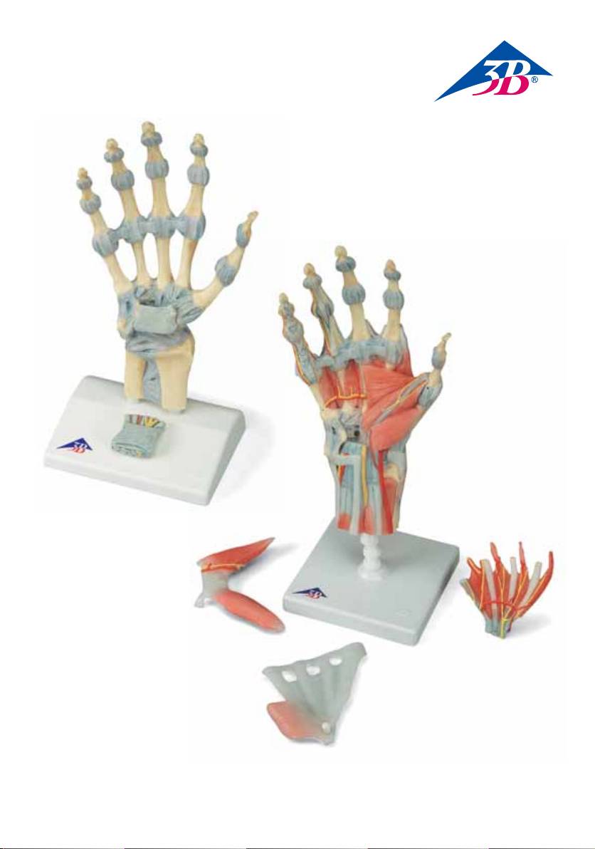
M33 - Hand Skeleton Model with Ligaments and Carpal Tunnel
This 3 part hand skeleton model shows the anatomical detail of the ligaments and tendons found in the
hand, wrist, and lower forearm. The interosseous membrane between the radius and ulna is shown along
with the bones of the hand. The flexor retunaculum is removable from the hand skeleton. In addition there
is a removable portion that can be fitted on the back of the hand model. This portion features the clinically
important structures of the carpal tunnel such as the flexor retinaculum, mediane nerve, and tendons of
the hand.
M33/1 - Hand Skeleton Model with Ligaments and Muscles
The bones, muscles, tendons, ligaments, nerves, arteries, and veins are all featured in this high quality 4
part model of the hand and lower forearm. The dorsal side of the hand shows the extensor muscles as well
as portions of the tendons at the wrist as they pass under the extensor retunaculum. The palmar face of
the hand is represented in three layers, the first two removable to allow detailed study of the deeper ana-
tomical layer of the hand. In additon clinically important structures such as the median nerve and superfi-
cial palmar arterial arch can be explored in detail in the hand model. The deepest anatomical layer allows
for study of the intrinsic muscles and deep palmar arterial arch in addition to other details of the anatomy
of the hand.
A Ligaments of the right hand, dorsal view
B Ligaments of the right hand, palmar view
C Carpal tunnel region of the right hand, palmar view
D Carpal tunnel insert, oblique view from the direction of the fingertips
E Ligaments of the right wrist, palmar view
F Dorsum of hand (dorsum manus) and extensor side of fingers of right hand
G Right palm of the hand (palma manus) with palmar aponeurosis
H Right palm of the hand (palma manus) without fasciae and palmar aponeurosis
I Superficial palmar arch of the right hand. Fasciae,
palmar aponeurosis and flexor retinaculum have been removed.
J Deep palmar arterial arch of the right hand
1 Palmar carpometacarpal ligaments
18 Distal phalanx
2 Tuberculum of trapezium bone
19 Proximal phalanx
3 Metacarpal bones I-V
20 Flexor retinaculum
4 Styloid process of radius
21 Interosseous membrane of forearm
5 Palmar radiocarpal ligament
22 Metacarpophalangeal joint, collateral
6 Radius
ligaments
7 Palmar radioulnar ligament
23 Deep transverse metacarpal ligament
8 Ulna
24 Middle phalanx
9 Palmar ulnocarpal ligament
25 Proximal interphalangeal joint
10 Tendon of flexor carpi ulnaris muscle
26 Distal interphalangeal joint
11 Pisiform bone
27 Distal interphalangeal joint, collateral
12 Hook of hamate bone
ligaments
13 Radiate carpal ligament
28 Proximal interphalangeal joint, collateral
14 Palmar metacarpal ligaments
ligaments
15 Pisometacarpal ligament
29 Dorsal metacarpal ligaments
16 Pisohamate ligament
30 Hamate bone
17 Palmar ligaments
31 Triquetrum bone
4
- 1
- 2
